Review
. 2010 Aug 18;29(16):2734-45.
doi: 10.1038/emboj.2010.170.

Cellular responses to extracellular guidance cues

Affiliations
Review

Cellular responses to extracellular guidance cues

Anastacia Berzat et al. EMBO J. .

Abstract

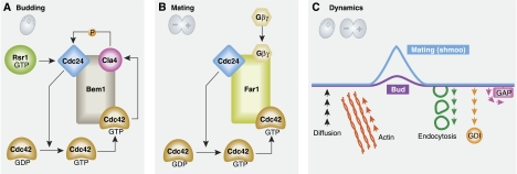
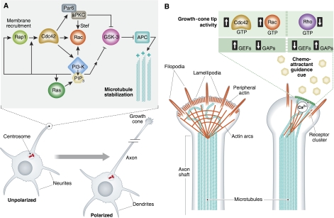
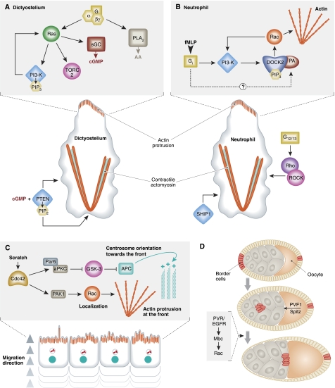
Extracellular guidance cues have a key role in orchestrating cell behaviour. They can take many forms, including soluble and cell-bound ligands (proteins, lipids, peptides or small molecules) and insoluble matrix substrates, but to act as guidance cues, they must be presented to the cell in a spatially restricted manner. Cells that recognize such cues respond by activating intracellular signal transduction pathways in a spatially restricted manner and convert the extracellular information into intracellular polarity. Although extracellular cues influence a broad range of cell polarity decisions, such as mitotic spindle orientation during asymmetric cell division, or the establishment of apical-basal polarity in epithelia, this review will focus specifically on guidance cues that promote cell migration (chemotaxis), or localized cell shape changes (chemotropism).


Conflict of interest statement

The authors declare that they have no conflict of interest.

Figures

Figure 1
Figure 1
Polarization in yeast. (A) Budding. Rsr1, a Ras-related small GTPase, localizes to the bud scar, a cortical remnant from the previous cell division. In its GTP-bound state, Rsr1 recruits Cdc24, an exchange factor for Cdc42. Activated Cdc42 captures the freely diffusing Bem1–Cdc24–Cla4 complex, through a direct interaction with Bem1, which serves to locally activate more Cdc42. There is evidence that Cla4 phosphorylates and activates Cdc24. This module serves as positive feedback loop creating a polarized patch of Cdc42 activity, which defines the new bud site. Subsequent recruitment of other Cdc42 targets and other Rho GTPases leads to the rearrangement of actin and microtubule cytoskeletons in the mother cell and to bud growth. (B) Mating. During mating, pheromone is recognized by surface receptors on cells of the opposite mating type. The pheromone gradient leads to the formation of a mating projection (shmoo) in the direction of the gradient source by first initiating receptor polarization, via an endocytosis and polarized recycling pathway. The subsequent release of Gβγ subunits from the associated heterotrimeric G protein recruits a scaffold protein, Far1, which is ordinarily resident in the nucleus in a complex with Cdc24. This forms the basis of a positive feedback loop. (C) Dynamics. The development of a single patch of active Cdc42 at the cortex is influenced by the rates of protein delivery to the membrane, either by free diffusion, or by transport along actin filaments, and by the rate of dissociation of proteins from the membrane, involving endocytosis, GDI-mediated solubilzation of Cdc42 and GAP stimulated GTP hydrolysis on Cdc42. The balance of these activities determines the overall distribution of the patch at the cortex to produce a round bud or a pointed shmoo.
Figure 2
Figure 2
Polarization and guidance in neurons. (A) Axon formation. The elongation of a single neurite to form an axon establishes a polarized neuron. In vivo this decision is guided by extracellular cues, but in vitro, stochastic changes in signalling activities at neurite tips, amplified by positive feedback loops, initiate axon formation. Several small GTPases localize to the tip of a presumptive axon. The proposed Rap1–Cdc42–Rac pathway is reminiscent of Rsr1–Cdc42–Rho in yeast polarization, although in the neurite, PI3 kinase seems to be the key downstream effector of Cdc42. It is capable of initiating a feedback loop through the generation of PIP3, a known activator of GEFs for Cdc42 and Rac. PI3 kinase is also a target of Ras and this too has been proposed to promote a feedback loop, probably through a Ras GEF. Changes in the dynamic behaviour of the actin and microtubule cytoskeletons at the neurite tip initiate axon extension. Rac directly regulates the actin and microtubule cytoskeletons through effector proteins such a LIM kinase and PAK kinase, but in addition the Par6–aPKC polarity complex, a direct target of Cdc42, localizes to the presumptive axon tip to promote stabilization of microtubules. This involves inhibition of GSK-3 leading to accumulation of APC at microtubule plus ends, though there is some uncertainty whether GSK-3 inhibition is direct (i.e. by phosphorylation) or indirect. (B) Axon guidance. A large number of extracellular cues are involved in guiding axons to their final destination. Both repulsive and attractive cues interact with the growth cone to influence the direction of elongation of axonal microtubules. They are thought to act by modifying the actin cytoskeleton in a spatially restricted manner within the growth cone causing it to turn towards or away from the source. In its simplest form, localized activation of Rho might be expected to lead to actomyosin-dependent retraction of the plasma membrane and induce turning away from a repellant, whereas activation of Rac or Cdc42 (↑) and inactivation of Rho (↓) could promote membrane protrusion and turning towards an attractant. Activation of GTPases can be achieved through activation of GEFs (↑) or inactivation of GAPs (↓). The situation is, however, clearly more complicated. Rac, for example, is activated during plexin-mediated repulsion, and the response to some repulsive cues can be reversed if the balance of cAMP and cGMP present within the growth cone is altered. Changes in cytosolic calcium seem to be essential for guidance signalling. A detailed understanding of these responses is lacking, but probably is dependent on the GEFs, GAPs and targets present in the growth cone of different neurons and their responsiveness to the different guidance cues encountered.
Figure 3
Figure 3
Directed cell migration. (A) Dictysotelium chemotaxis. Ras is activated in response to a gradient of cAMP interacting with a surface GPCR. The receptor and associated G protein, as well as Ras, remain evenly distributed around the cell, but active, GTP-bound Ras is localized specifically in the direction of the gradient. Ras activates PI3 kinase to produce PIP3, which leads not only to the recruitment of proteins involved in modifying the actin cytoskeleton, such as SCAR/WAVE and Rac, but also a Ras GEF, which could potentially generate a positive feedback loop. PTEN localizes at the sides and rear of the cell to spatially restrict the distribution of PIP3 within the plasma membrane. Two other second messenger systems have important roles in directional sensing, PLA2 and soluble guanylyl cyclase (sGC), which produce arachidonic acid metabolites (AA) and cGMP, respectively. Contractile actomyosin forces at the sides and rear together with actin-driven protrusions at the front drive migration. (B) Neutrophil chemotaxis. An extracellular gradient of the bacterial-derived peptide fMLP, interacting with a GPCR (FMP2), leads to activation of an associated Gi and PI3 kinase. The PIP3 produced recruits DOCK2 to activate Rac, which can activate PI3 kinase and set up a positive feedback loop. Sustained recruitment of DOCK2 also requires localized generation of phosphatidic acid (PA) through activation of PLD. Another G protein, G12/13, activates Rho and ROCK at the rear of the cell to promote actomyosin contractility. The lipid phosphatase SHIP1 is required to maintain spatially localized PIP3. (C) Collective migration in vitro. After scratching a monolayer of primary embryo-derived fibroblasts, cells migrate into the free space collectively as a sheet. Cell–cell contacts (N-cadherin) are maintained during migration. Cdc42 is activated at the leading edge of front row cells and, via PAK kinase, serves to localize Rac activity such that actin polymerization and protrusive activity are restricted to the front of the migrating cell. Through another effector, the Par6–aPKC complex, Cdc42 causes the microtubule cytoskeleton to reorganize such that the centrosome and Golgi face the direction of migration. Par6–aPKC leads to the inhibition of GSK-3 specifically at the front of the cell, through a pathway that involves Wnt5a and Disheveled (data not shown), and causes the tumour suppressor protein APC to associate with and stabilize the plus ends of neighbouring microtubules. (D) Collective migration in vivo. Border cells (yellow) in the Drosophila ovary delaminate from the follicular epithelium (brown) and migrate as a group through nurse cells (white/grey) towards the oocyte. PVR and EGFR receptors in the border cells recognize a gradient of extracellular cues (PVF1 and Spitz) from the oocyte, and activate Rac through the Rac GEF, Mbc, to induce forward facing protrusion in cells at the front of the group. Cell–cell interactions (through E-cadherin) are required to be maintained throughout migration, though cells continuously reorganize within the group.

Similar articles

Cited by

References

    1. Adler CE, Fetter RD, Bargmann CI (2006) UNC-6/Netrin induces neuronal asymmetry and defines the site of axon formation. Nat Neurosci 9: 511–518 - PMC - PubMed
    1. Aman A, Piotrowski T (2010) Cell migration during morphogenesis. Dev Biol 341: 20–33 - PubMed
    1. Andrew N, Insall RH (2007) Chemotaxis in shallow gradients is mediated independently of PtdIns 3-kinase by biased choices between random protrusions. Nat Cell Biol 9: 193–200 - PubMed
    1. Arimura N, Inagaki N, Chihara K, Menager C, Nakamura N, Amano M, Iwamatsu A, Goshima Y, Kaibuchi K (2000) Phosphorylation of collapsin response mediator protein-2 by Rho-kinase. Evidence for two separate signaling pathways for growth cone collapse. J Biol Chem 275: 23973–23980 - PubMed
    1. Barberis D, Artigiani S, Casazza A, Corso S, Giordano S, Love CA, Jones EY, Comoglio PM, Tamagnone L (2004) Plexin signaling hampers integrin-based adhesion, leading to Rho-kinase independent cell rounding, and inhibiting lamellipodia extension and cell motility. FASEB J 18: 592–594 - PubMed

Publication types