. 2011 Jan;19(1):30-5.
doi: 10.1038/ejhg.2010.140. Epub 2010 Aug 18.

Alternative splice variants of the USH3A gene Clarin 1 (CLRN1)

Affiliations

Alternative splice variants of the USH3A gene Clarin 1 (CLRN1)

Hanna Västinsalo et al. Eur J Hum Genet. 2011 Jan.

Abstract

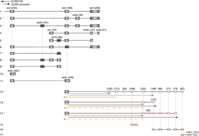
Clarin 1 (CLRN1) is a four-transmembrane protein expressed in cochlear hair cells and neural retina, and when mutated it causes Usher syndrome type 3 (USH3). The main human splice variant of CLRN1 is composed of three exons that code for a 232-aa protein. In this study, we aimed to refine the structure of CLRN1 by an examination of transcript splice variants and promoter regions. Analysis of human retinal cDNA revealed 11 CLRN1 splice variants, of which 5 have not been previously reported. We studied the regulation of gene expression by several promoter domains using a luciferase assay, and identified 1000 nt upstream of the translation start site of the primary CLRN1 splice variant as the principal promoter region. Our results suggest that the CLRN1 gene is significantly more complex than previously described. The complexity of the CLRN1 gene and the identification of multiple splice variants may partially explain why mutations in CLRN1 result in substantial variation in clinical phenotype.


Figures

Figure 1
Figure 1
Known and novel splice variants of CLRN1. (1) NM_174878, the main CLRN1 variant comprises exons 0, 2, and 3. The 5′ UTR and 3′ UTRs are not counted in the exon nucleotide number (in brackets). Known human CLRN1 splice variants or EST sequences from gene databanks: (4) NM_052995, the original CLRN1 variant with exons 1, 2, 3a, and 3b reported in 2001; (5) AF388368, coding region only 30-aa long; (6) BM666773 found from retina, exons 1 and 2 are coded in an alternative reading frame compared with the other splice variants, and a stop codon appears in this frame in exon 2 (presence of a stop codon marked with a red star); (10) BX491536, exon 0 open reading frame (ORF) continues to the intron until stop codon, potentially codes for 87-aa protein; and (11) CV570593, exon 2 continues to the intron, codes for a potentially 167-aa long protein. Novel splice variants found in human retina cDNA library (marked with an asterisk): (2) HM626132, main variant with added exon 2b; (3) HM626133, main variant with added exons 0b and 2b; and (7–9) HM626134, HM626135 and HM626136, splice variants between exons 0 and 1b. CLRN1 exon 2 is also connected by splicing to three EST sequences downstream of CLRN1. Downstream EST sequences from databanks are (15) BE673203, (16) DV080481, and (17) DV080691. The 5′ UTR is unknown, but presumed in this figure to start from exon 2. Possible ORFs in these variants (12–14: HM626137, HM626138, and HM626139) are depicted as arrows. ORFs that continue the same ORF as in CLRN1 exon 2 are depicted as green arrows, reading frames in blue and red begin from exon 2, but are not in the same ORF as CLRN1. Two ORFs running in opposite direction than CLRN1 ORF are depicted as orange in either solid or dashed line. The exon and intron sizes are not drawn in scale.
Figure 2
Figure 2
Potential sites of gene expression regulation in the proximal 1550 nt of each of CLRN1's primary exons.
Figure 3
Figure 3
CLRN1 promoter region activity levels. Possible promoter regions were inserted in the pGLuc expression vector and transfected in triplicate into BHK cells. The luciferase activity was measured from the following constructs (upstream of translation start site or exon splice site): (1) exon 0: 1–500 nt, (2) exon 0: 1–1000 nt, (3) exon 0: 1–1550 nt, (4) exon 1: 1–1550 nt, (5) exon 2: 1–1550 nt, (6) exon 3: 1–1550 nt, and (7) unmodified pGluc vector. The relative activity levels were set to positive control pCMV-Gluc vector (New England BioLabs) as 100% and untransfected cell culture media signal level was subtracted from these values. Error bars reflect 1 SD. Promoter region ClustalW2 scores (sequence conservation between mouse and human) are displayed as an insert: (a) 500 nt upstream exon 0, (b) 500–1000 nt upstream exon 0, (c) 1000–1550 nt upstream exon 0, (d) 1–1550 nt upstream exon 0, (e) 1–1550 nt upstream exon 1, (f) 1–1550 nt upstream exon 2, and (g) 1–1550 nt upstream exon 3.

References

    1. Spandau UH, Rohrschneider K. Prevalence and geographical distribution of Usher syndrome in Germany. Graefes Arch Clin Exp Ophthalmol. 2002;240:495–498. - PubMed
    1. Saihan Z, Webster AR, Luxon L, Bitner-Glindzicz M. Update on Usher syndrome. Curr Opin Neurol. 2009;22:19–27. - PubMed
    1. Pakarinen L, Tuppurainen K, Laippala P, Mäntyjärvi M, Puhakka H. The ophthalmological course of Usher syndrome type III. Int Ophthalmol. 1995;19:307–311. - PubMed
    1. Ness SL, Ben-Yosef T, Bar-Lev A, et al. Genetic homogeneity and phenotypic variability among Ashkenazi Jews with Usher syndrome type III. J Med Genet. 2003;40:767–772. - PMC - PubMed
    1. Cohen M, Bitner-Glindzicz M, Luxon L. The changing face of Usher syndrome: clinical implications. Int J Audiol. 2007;46:82–93. - PubMed

Publication types