. 2010 May 21:3:12.
doi: 10.3389/fnmol.2010.00012. eCollection 2010.

Characterization of the Brain 26S Proteasome and its Interacting Proteins

Affiliations

Characterization of the Brain 26S Proteasome and its Interacting Proteins

Hwan-Ching Tai et al. Front Mol Neurosci. .

Abstract

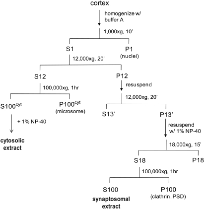
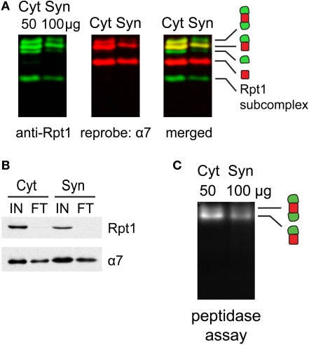
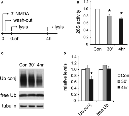
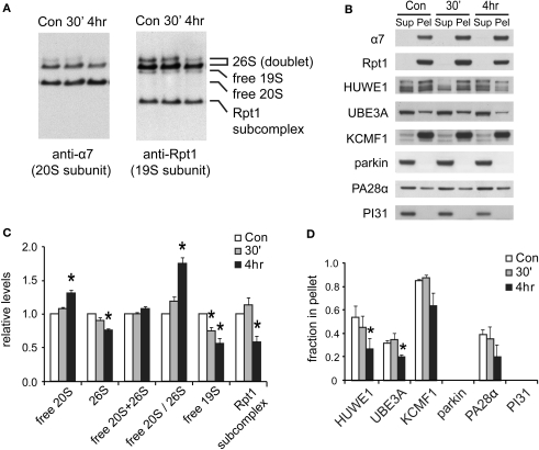
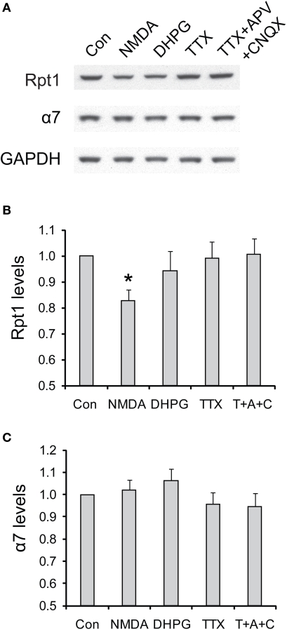
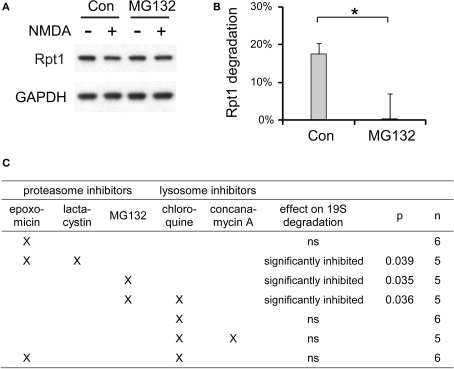
Proteasome-mediated proteolysis is important for synaptic plasticity, neuronal development, protein quality control, and many other processes in neurons. To define proteasome composition in brain, we affinity purified 26S proteasomes from cytosolic and synaptic compartments of the rat cortex. Using tandem mass spectrometry, we identified the standard 26S subunits and a set of 28 proteasome-interacting proteins that associated substoichiometrically and may serve as regulators or cofactors. This set differed from those in other tissues and we also found several proteins that associated only with either the cytosolic or the synaptic proteasome. The latter included the ubiquitin-binding factor TAX1BP1 and synaptic vesicle protein SNAP-25. Native gel electrophoresis revealed a higher proportion of doubly-capped 26S proteasome (19S-20S-19S) in the cortex than in the liver or kidney. To investigate the interplay between proteasome regulation and synaptic plasticity, we exposed cultured neurons to glutamate receptor agonist NMDA. Within 4 h, this agent caused a prolonged decrease in the activity of the ubiquitin-proteasome system as shown by disassembly of 26S proteasomes, decrease in ubiquitin-protein conjugates, and dissociation of the ubiquitin ligases UBE3A (E6-AP) and HUWE1 from the proteasome. Surprisingly, the regulatory 19S particles were rapidly degraded by proteasomal, not lysosomal degradation, and the dissociated E3 enzymes also degraded. Thus the content of proteasomes and their set of associated proteins can be altered by neuronal activity, in a manner likely to influence synaptic plasticity and learning.

Keywords: NMDA; UBE3A; proteasome; proteasome-interacting protein; synaptic plasticity; ubiquitin ligase.


Figures

Figure 1
Figure 1
A schematic showing the preparation of synaptosomal and cytosolic extracts from rat cortices. We include an additional centrifugation step (1 h, 100,000 × g) after solubilization of synaptosomes with NP-40 and before incubation with the UBL-domain. Solubilization leads to binding of an unrelated clathrin complex to the UBL matrix, which complicates the mass spectrometric analysis (data not shown). The respective centrifugation step removes the bulk of this clathrin complex while the proteasome remains fully soluble.
Figure 2
Figure 2
(A) Proteasome species found in different cell types. Extracts of HeLa cell (30 μg), rat cortex (60 μg), rat liver (40 μg) and rat kidney (50 μg) were resolved by 2–5% gradient native gel, immunoblotted against 19S subunit Rpt1, and reprobed against 20S subunit α7. Protein bands were pseudo-colored and merged to illustrate the presence of proteasomes and related species (from top to bottom): doubly-capped 26S proteasome, singly-capped 26S proteasome, free 19S particle and free 20S particle. A fast-migrating band corresponding to a subcomplex of the 19S containing Rpt1 protein was also observed. (B) The distribution of different proteasome species in (A), quantified by densitometry (mean ± SEM, n = 6). The experiment was performed independently six times with tissues from three animals. By one-way ANOVA and Turkey's multiple comparison test, doubly-capped 26S levels in cortex are found to be significantly higher (p < 0.01) than those in liver, kidney or HeLa cells.
Figure 3
Figure 3
(A) Proteasome integrity after subcellular fractionation. Cytosolic (Cyt) and synaptic (Syn) extracts prepared from rat cortices were resolved by 2–5% gradient native gel, immunoblotted against 19S subunit Rpt1, and reprobed for 20S subunit α7. Singly-, doubly-capped 26S, 20S proteasomes and free 19S were detected in both fractions. (B) Efficacy of 26S proteasome isolation by UBL affinity chromatography. Cytosolic (Cyt) and synaptic (Syn) extracts were incubated with GST-UBL and glutathione sepharose. The input (IN) and flow-through (FT) materials were analyzed by SDS-PAGE and immunoblotted against Rpt1 and α7. The depletion of Rpt1 from the flow-through indicated that most 26S proteasomes and 19S particles remained intact during subcellular fractionation and were captured with high efficiency. The high remaining level of α7 subunit in the flow-through is consistent with the abundance of free 20S particles detected in (A). (C) Cytosolic and synaptosomal extracts contain active 26S proteasomes. Cytosolic (Cyt) and synaptic (Syn) extracts isolated from rat cortices were resolved by 2–5% gradient native gel, followed by incubation with Suc-LLVY-AMC, a fluorogenic proteasome substrate. Active 26S proteasomes appear as fluorescent bands, corresponding to doubly- and singly-capped 26S proteasomes.
Figure 4
Figure 4
(A) Purity of isolated 26S proteasomes. Cytosolic (Cyt) and synaptic (Syn) 26S proteasomes were isolated using GST-UBL/His-UIM. In control (Con) experiments, GST was added instead of GST-UBL. Samples were resolved by 3–8% gradient native gel and silver-stained. (B) The same samples in (A) analyzed by SDS-PAGE, followed by silver staining. The control experiments showed only a few non-specific protein bands. (C) Activity of isolated proteasomes. Chymotrypsin-like peptidase activity of isolated 26S proteasomes resolved by 3–8% native gel was assayed with Suc-LLVY-AMC. (D) Differences in capped proteasome ratios. Silver-stained 26S proteasome bands in (C) were quantified by densitometry. The ratio of doubly- over singly-capped 26S was significantly higher in the cytosol (Cyt) than the synaptosome (Syn) (mean ± SEM, n = 4, *p < 0.05 by paired t-test).
Figure 5
Figure 5
(A) Verification of drebrin and GRASP-1 as interactors of synaptic proteasomes. Both proteins were initially detected in synaptic proteasomes by mass spectrometry, based on a single matching peptide (Table 2). These specific interactions are confirmed by SDS-PAGE and Western blotting. Synaptosome (Syn)-derived proteasomes (26S) and control (Con) purifications using only GST are probed with specific antibodies against drebrin, GRASP-1, and 19S subunit Rpt1. (B) The VCP complex does not interact appreciably with GST-UBL. 26S proteasomes from the cytosolic (Cyt) and synaptic (Syn) extracts were purified by the GST-UBL/His-UIM method. The input (IN) material and purified 26S proteasomes were immunoblotted against VCP and Rpt1. The amount of VCP captured by the GST-UBL matrix was undetectable by Western blot in three independent experiments. (C) Verification of proteasome-interacting proteins by co-sedimentation. Cytosolic (Cyt) and synaptosomal (Syn) extracts were subjected to centrifugation at 100,000 × g for 6 h. The supernatant (Sup) and pellet (Pel) materials were loaded at 1:1 ratio and resolved by SDS-PAGE. The sedimentation of proteasomes was confirmed by the high levels of 20S subunit α7 and 19S subunit Rpt1 in the pellet fraction. Proteins identified by mass spectrometry in 26S proteasomes (26S MS) from the cytosol and the synaptosome are indicated by the positive sign. Agreement between mass spectrometry data and co-sedimentation data was observed for all seven 26S-interacting proteins (UBE3A, HUWE1, KCMF1, USP14, PSMD9, ECM29, 14-3-3γ) probed by specific antibodies (see Materials and Methods). Parkin and PI31 did not show appreciable association with brain proteasomes. Results shown here are representative of three experiments.
Figure 6
Figure 6
UPS activity is reduced after NMDA treatment. (A) Cultured hippocampal neurons were treated with 20 μM NMDA for 3 min, a standard procedure for inducing chemical LTD. Whole-cell lysates were prepared at 30 min or 4 h post-treatment. (B) Decreases in proteasome activity after NMDA exposure. 26S proteasome activities in neuronal lysates were assayed with Suc-LLVY-AMC (see Materials and Methods). NMDA-treated samples (30’ and 4 h) are normalized to the untreated control (Con) (mean ± SEM, n = 5, *p < 0.05 by paired t-test). (C) Ubiquitin conjugates decrease after NMDA exposure. Neuronal lysates resolved by SDS-PAGE were blotted using antibodies against ubiquitin-protein conjugates (Ub conj) and free monomeric ubiquitin (free Ub). Tubulin served as the loading control. (D) Signals in (C) quantified by densitometry (n = 5, *p < 0.05 by paired t-test).
Figure 7
Figure 7
Changes in proteasome complexes after NMDA exposure. (A) The disassembly of 26S proteasomes. Neuronal lysates collected at 30’ and 4 h post-NMDA (20 μM, 3 min) were resolved by 2–5% gradient native gel and immunoblotted against 20S subunit α7 and 19S subunit Rpt1. (B) Changes in proteasome-associated proteins. Lysates from (A) were subjected to ultracentrifugation to sediment proteasomes. Equal amounts of supernatant (Sup) and pellet (Pel) materials were analyzed by SDS-PAGE. The sedimentation property of proteasome-interacting proteins (HUWE1, UBE3A, KCMF1, PA28α) was examined by Western blotting. Parkin and PI31 were not detected in the pellet. (C) Quantification of changes in proteasome distributions in (A) by densitometry (n = 6, mean ± SEM, *p < 0.05 by paired t-test). (D) The graph represents the sedimentation data in (B). The protein level in the pellet is divided by the total level (supernatant + pellet), and the results are plotted (n = 4, *p < 0.05 by paired t-test).
Figure 8
Figure 8
Degradation of proteasome components after NMDA exposure. (A) The degradation of 19S subunits. Neuronal lysates collected at 30’ and 4 h post-NMDA were resolved by SDS-PAGE and probed for total levels of 20S subunit α7, 19S subunit Rpt1, and VCP (an abundant UPS protein). Tubulin served as the loading control. (B) Quantification of the results in (A) by densitometry (n = 6, mean ± SEM, *p < 0.05 by paired t-test). (C) Degradation of proteasome-interacting E3s. Neuronal lysates from (A) were immunoblotted against E3s (HUWE1, UBE3A, KCMF1) and DUBs (USP14) that interact with proteasomes. (D) The graph represents protein levels in (C) normalized to α7 levels (n = 5, *p < 0.05 by paired t-test).
Figure 9
Figure 9
The degradation of 19S particles is specific to NMDA treatment. (A) Cultured hippocampal neurons were briefly treated with NMDA (20 μM, 3 min) or DHPG (100 μM, 10 min), or persistently treated with TTX (2 μM) or TTX + CNQX + APV (2, 40, and 50 μM, respectively). Only the vehicle was added in control experiments (Con). Neuronal lysates were collected at 4 h after drug addition, and analyzed by SDS-PAGE and Western blot for the levels of 19S subunit Rpt1 and 20S subunit α7. GAPDH served as loading control. (B,C) represent the results in (A) quantified by densitometry. Rpt1 and α7 levels (mean ± SEM) were normalized to the control. Statistical significance (*p < 0.05) was determined by paired t-test against the control (n = 8 for NMDA and DHPG, n = 4 for TTX and TTX + APV + CNQX).
Figure 10
Figure 10
The degradation of 19S particles is proteaosme-dependent. (A) Proteasome inhibitor MG132 (40 μM) was added to cultured hippocampal neurons 30 min before NMDA stimulation (20 μM, 3 min). After NMDA washed-out, MG132 was applied again for 4 h before neuronal lysates were collected. In control (Con) experiments, neurons were only treated with NMDA. Rpt1 levels were determined by SDS-PAGE and immunoblotting. GAPDH served as loading control. (B) 19S degradation induced by NMDA in (A) was quantified by densitometry (n = 5, mean ± SEM). Statistical significance (*p < 0.05) was determined by t-test assuming unequal variance. (C) The same experiments as in (A) and (B), performed with different combinations of proteasome inhibitors (2 μM epoxomicin; 10 μM lactacystin; 40 μM MG132) and lysosome inhibitors (150 μM chloroquine; 100 nM concanamycin A). Statistical significance was determined using t-test (p-value is listed, or ns for not significant, p > 0.05).

Similar articles

Cited by

References

    1. Aakalu G., Smith W. B., Nguyen N., Jiang C., Schuman E. M. (2001). Dynamic visualization of local protein synthesis in hippocampal neurons. Neuron 30, 489–50210.1016/S0896-6273(01)00295-1 - DOI - PubMed
    1. Aguilaniu H., Gustafsson L., Rigoulet M., Nystrom T. (2003). Asymmetric inheritance of oxidatively damaged proteins during cytokinesis. Science 299, 1751–175310.1126/science.1080418 - DOI - PubMed
    1. Anderson C., Crimmins S., Wilson J. A., Korbel G. A., Ploegh H. L., Wilson S. M. (2005). Loss of Usp14 results in reduced levels of ubiquitin in ataxia mice. J. Neurochem. 95, 724–73110.1111/j.1471-4159.2005.03409.x - DOI - PubMed
    1. Bajorek M., Finley D., Glickman M. H. (2003). Proteasome disassembly and downregulation is correlated with viability during stationary phase. Curr. Biol. 13, 1140–114410.1016/S0960-9822(03)00417-2 - DOI - PubMed
    1. Bedford L., Hay D., Devoy A., Paine S., Powe D. G., Seth R., Gray T., Topham I., Fone K., Rezvani N., Mee M., Soane T., Layfield R., Sheppard P. W., Ebendal T., Usoskin D., Lowe J., Mayer R. J. (2008a). Depletion of 26S proteasomes in mouse brain neurons causes neurodegeneration and Lewy-like inclusions resembling human pale bodies. J. Neurosci. 28, 8189–819810.1523/JNEUROSCI.2218-08.2008 - DOI - PMC - PubMed