

Chordoid glioma : a case report of unusual location and neuroradiological characteristics
- PMID: 20717514
- PMCID: PMC2916150
- DOI: 10.3340/jkns.2010.48.1.62
Chordoid glioma : a case report of unusual location and neuroradiological characteristics
Abstract
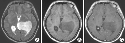
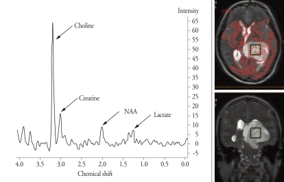
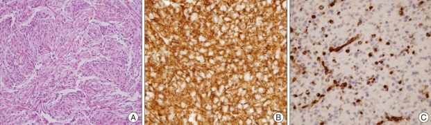
Since the World Health Organization (WHO) classification for central nervous system neoplasms was declared in 2000, chordoid glioma of the third ventricle has been noted as a newly recognized tumor for central nervous system neoplasms. Although there is not enough universal experience to know the nature of this tumor due to its rarity, the origin of chordoid glioma was guardedly proposed to be the ependymal cells of the third ventricle. Such an idea has been primarily based on the specific location of the tumor, that is, third ventricle, suprasellae, and hypothalamus. However, we report a rare case of histologically confirmed chordoid glioma located in the left thalamus, not attached to any of the midline structures having unusual neuroradiological characteristics.
Keywords: Chordoid glioma; Radiological feature; Third ventricle.
Figures
Similar articles
-
Chordoid glioma of the third ventricle: four cases including one case with papillary features.Neuropathology. 2013 Apr;33(2):134-9. doi: 10.1111/j.1440-1789.2012.01333.x. Epub 2012 Jun 21. Neuropathology. 2013. PMID: 22716306
-
Chordoid Glioma with Psychosis: Case Report.P R Health Sci J. 2018 Sep;37(3):174-176. P R Health Sci J. 2018. PMID: 30188563
-
Chordoid glioma: report of two rare examples with unusual features.Acta Neurochir (Wien). 2008 Mar;150(3):295-300; discussion 300. doi: 10.1007/s00701-008-1420-x. Epub 2008 Feb 4. Acta Neurochir (Wien). 2008. PMID: 18246456
-
Chordoid glioma of the third ventricle: a report of two new cases, with further evidence supporting an ependymal differentiation, and review of the literature.Am J Surg Pathol. 2002 Oct;26(10):1330-42. doi: 10.1097/00000478-200210000-00010. Am J Surg Pathol. 2002. PMID: 12360048 Review.
-
Chordoid glioma of the third ventricle: a patient presenting with SIADH and a review of this rare tumor.Pituitary. 2016 Aug;19(4):356-61. doi: 10.1007/s11102-016-0711-8. Pituitary. 2016. PMID: 26879322 Review.
Cited by
-
Chordoid glioma in the thalamus of a child: Rare location and atypical imaging findings.BJR Case Rep. 2021 Jan 8;7(3):20200108. doi: 10.1259/bjrcr.20200108. eCollection 2021 May 1. BJR Case Rep. 2021. PMID: 34131491 Free PMC article.
-
A 29-year-old man with progressive short term memory loss.Brain Pathol. 2014 Jan;24(1):103-6. doi: 10.1111/bpa.12107. Brain Pathol. 2014. PMID: 24345225 Free PMC article. No abstract available.
-
One of a kind-chordoid glioma in the fourth ventricle: a case report and literature review.Acta Radiol Open. 2020 Dec 14;9(12):2058460120980143. doi: 10.1177/2058460120980143. eCollection 2020 Dec. Acta Radiol Open. 2020. PMID: 33403125 Free PMC article.
-
Prognostic factors for recurrence and complications in the surgical management of primary chordoid gliomas: A systematic review of literature.Clin Neurol Neurosurg. 2015 Nov;138:129-36. doi: 10.1016/j.clineuro.2015.08.011. Epub 2015 Aug 19. Clin Neurol Neurosurg. 2015. PMID: 26342205 Free PMC article.
-
Occurrence of Chordoid Glioma With Sodium Ion Metabolism Disorder 5 Years After Meningioma Surgery and Whole-Exome Sequencing: A Case Report and Literature Review.Front Genet. 2021 May 10;12:617575. doi: 10.3389/fgene.2021.617575. eCollection 2021. Front Genet. 2021. PMID: 34040630 Free PMC article.
References
-
- Baehring JM, Bannykh S. Chordoid glioma of the third ventricle. J Neurooncol. 2006;76:269. - PubMed
-
- Brat DJ, Scheithauer BW, Staugaitis SM, Cortez SC, Brecher K, Burger PC. Third ventricular chordoid glioma : a distinct clinicopathologic entity. J Neuropathol Exp Neurol. 1998;57:283–290. - PubMed
-
- Buccoliero AM, Caldarella A, Gallina P, Di Lorenzo N, Taddei A, Taddei GL. Chordoid glioma : clinicopathologic profile and differential diagnosis of an uncommon tumor. Arch Pathol Lab Med. 2004;128:e141–e145. - PubMed
-
- Carrasco R, Pascual JM, Reina T, Nieto S, Linera J, Sola RG. Chordoid glioma of the third ventricle attached to the optic chiasm. Successful removal through a trans-lamina terminalis approach. Clin Neurol Neurosurg. 2008;110:828–833. - PubMed
-
- Castellano-Sanchez AA, Recine MA, Restrepo R, Howard LH, Robinson MJ. Chordoid glioma : a novel tumor of the third ventricle. Ann Diagn Pathol. 2000;4:373–378. - PubMed
Publication types
LinkOut - more resources
Full Text Sources
