. 2010 Sep;2(9):424-34.
doi: 10.1039/c0ib00040j. Epub 2010 Aug 17.

Endogenous patterns of mechanical stress are required for branching morphogenesis

Affiliations

Endogenous patterns of mechanical stress are required for branching morphogenesis

Nikolce Gjorevski et al. Integr Biol (Camb). 2010 Sep.

Abstract

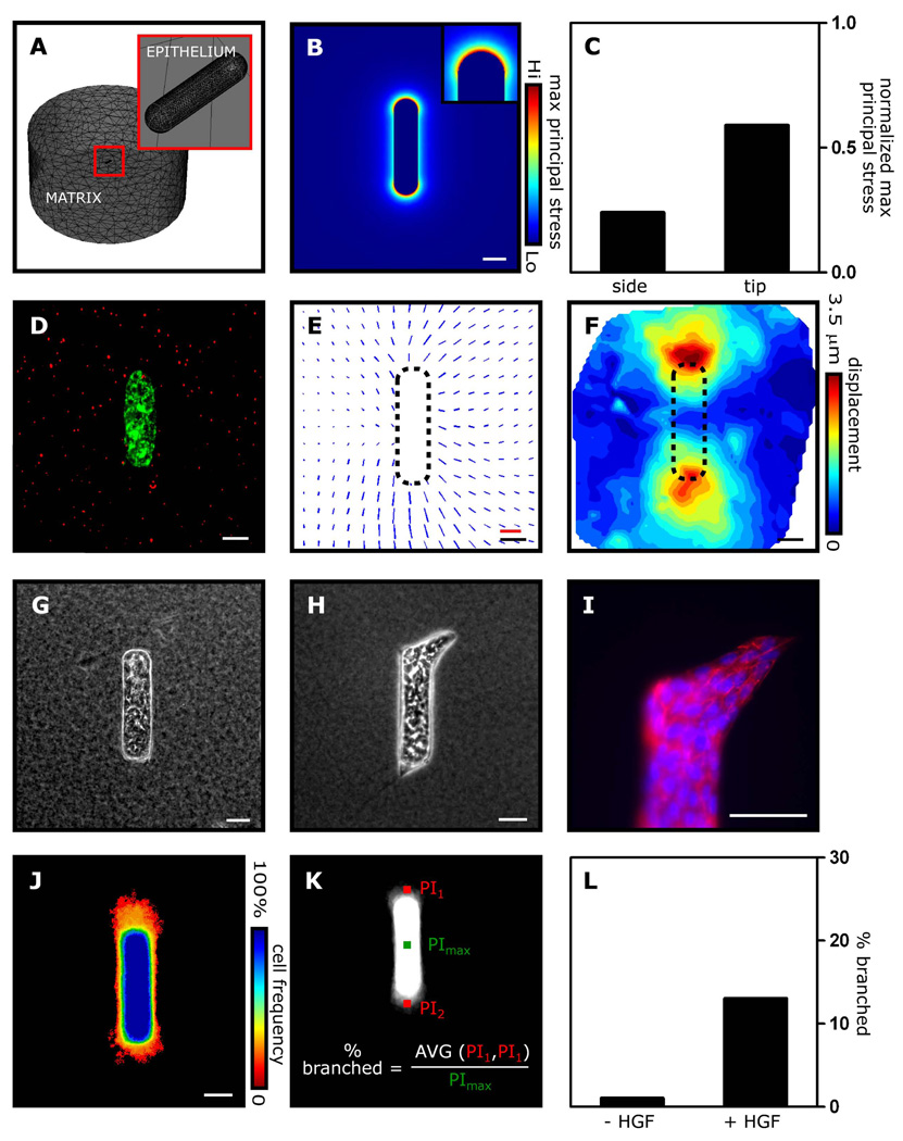
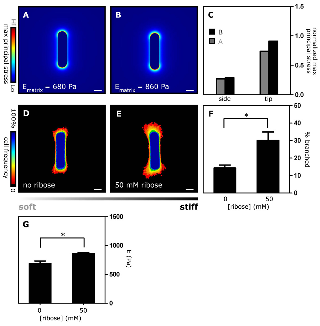
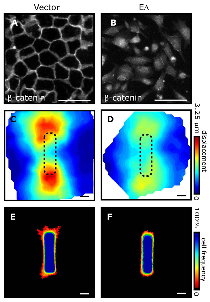
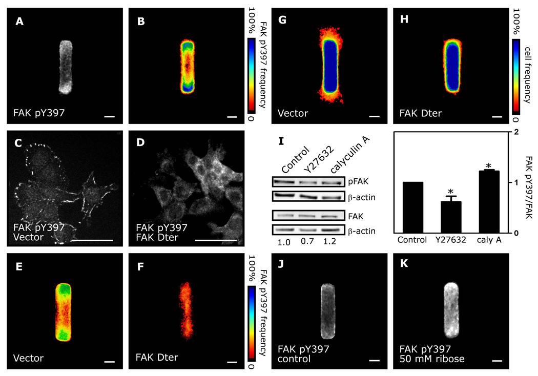
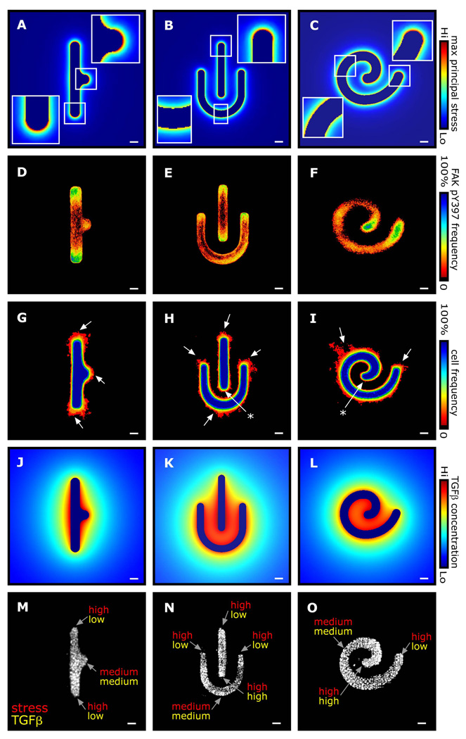
Spatial patterning of cell behaviors establishes the regional differences within tissues that collectively develop branched organs into their characteristic treelike shapes. Here we show that the pattern of branching morphogenesis of three-dimensional (3D) engineered epithelial tissues is controlled in part by gradients of endogenous mechanical stress. We used microfabrication to build model mammary epithelial tissues of defined geometry that branched in a stereotyped pattern when induced with growth factors. Branches initiated from sites of high mechanical stress within the tissues, as predicted numerically and measured directly using 3D traction force microscopy. Branch sites were defined by activation of focal adhesion kinase (FAK), inhibition of which disrupted morphogenesis. Stress, FAK activation, and branching were all altered by manipulating cellular contractility, matrix stiffness, intercellular cohesion and tissue geometry. These data suggest that the pattern and magnitude of mechanical stress across epithelial tissues cooperate with biochemical signals to specify branching pattern.


Figures

Fig. 1
Fig. 1. Patterns of mechanical stress correspond to sites of branching in epithelial tissues
(A) FEM mesh, showing the cellular (epithelium) and ECM (matrix) portions of the stress model. (B, C) FEM stress profile of an epithelial tubule. (D) Fluorescent image of engineered mammary epithelial tubule showing cellular membranes (green) and microbeads (red) embedded in the surrounding collagen gel. (E) Map of bead displacements around a single engineered tubule and (F) average displacements around 15 tubules. (G) Phase contrast image of engineered tubule before induction of branching. (H) Phase contrast image, (I) fluorescent image of E-cadherin (red) and nuclei (blue), and (J) frequency map of engineered tubules after induction of branching. (K, L) Branching is quantified by measuring the pixel intensity (PI) at a fixed location away from the tip in the grayscale frequency map, and then calculating the percent of tubules that have extended branches. Scale bars, 50 µm; displacement scale bar (red), 3 µm.
Fig. 2
Fig. 2. Patterns of mechanical stress and branching morphogenesis are affected by cellular contractility
Mechanical stress profiles of tubules with increasing contractility: prestress (pt) that yields prestrain (ε0) of (A) 0.2 % (B) 0.6 % (C) 1% and (D) combined stress profiles. (E–H) Matrix displacement maps around control tubules and tubules treated with 10 µM Y27632 or 0.1 nM calyculin A. (I–K) Frequency maps of branching from control tubules and tubules treated with 10 µM Y27632 or 0.1 nM calyculin A. (L) Branching was also quantified by measuring the pixel intensity 12 µm away from the tubule tip. Branch frequency decreases when tubules are treated with Y27632, and increases when tubules are treated with calyculin A. (M–O) Frequency maps and (P) quantification of branching from tubules constructed of cells transduced with adenovirus encoding RhoA-N19, RhoA-L63, or control vector. *p<0.05; **p<0.01; Scale bars, 50 µm.
Fig. 3
Fig. 3. Patterns of mechanical stress and branching morphogenesis are affected by matrix stiffness
Mechanical stress profiles of tubules surrounded by matrices of different stiffness: (A) Ematrix = 680 Pa, (B) Ematrix = 860 Pa and (C) combined stress profiles. Frequency maps of branching tubules embedded in collagen gel crosslinked with (D) 0 and (E) 50 mM D-ribose. (F) Branch frequency increases as the stiffness of the collagen gel increases (G). Rheological measurements of collagen gels crosslinked with D-ribose. *p<0.05; Scale bars, 50 µm.
Fig. 4
Fig. 4. Intercellular cohesion is required for patterning stress and branching morphogenesis
β-catenin immunofluorescence in (A) control mammary epithelial cells and (B) cells expressing EΔ. Matrix displacement maps following relaxation of (C) control tubules and (D) tubules expressing EΔ. Frequency maps of branching from (E) control tubules and (F) tubules expressing EΔ. Scale bars, 50 µm.
Fig. 5
Fig. 5. FAK is activated at tips of tubules and required for branching morphogenesis
(A) FAK pY397 immunofluorescence and (B) frequency map in engineered tubules. FAK pY397 immunofluorescence in (C) control cells and (D) cells expressing FAK Dter. Frequency maps of FAK pY397 in (E) control tubules and (F) tubules expressing FAK Dter. Frequency map of branching from (G) control tubules and (H) tubules expressing FAK Dter. (I) Western blot analysis of FAK pY397 and total FAK in tubules treated with Y27632 or calyculin A. FAK pY397 immunofluorescence in (J) control tubules and (K) tubules constructed in collagen crosslinked using ribose. *p<0.05; Scale bars, 50 µm.
Fig. 6
Fig. 6. Patterns of mechanical stress and branching morphogenesis are affected by tissue geometry
(A–C) FEM stress profiles of tubules of various geometry. Frequency maps of (D–F) FAK pY397 immunofluorescence and (G–I) branching from tubules of the corresponding geometries. Arrows denote regions of high mechanical stress that branch, and asterisks denote regions of high mechanical stress that do not branch. (J–L) Predicted concentration profiles of TGFβ around tubules of various geometry. (M–O) Relative strength of the mechanical and biochemical inputs as a function of position within the tissue. Scale bars, 50 µm.

Similar articles

Cited by

References

    1. Affolter M, Bellusci S, Itoh N, Shilo B, Thiery JP, Werb Z. Tube or not tube: remodeling epithelial tissues by branching morphogenesis. Dev. Cell. 2003;4:11–18. - PubMed
    1. Davies JA. Branching morphogenesis. New York: Landes Bioscience; 2006.
    1. Affolter M, Zeller R, Caussinus E. Tissue remodelling through branching morphogenesis. Nat. Rev. Mol. Cell Biol. 2009;10:831–842. - PubMed
    1. Griffith LG, Swartz MA. Capturing complex 3D tissue physiology in vitro. Nat. Rev. Mol. Cell Biol. 2006;7:211–224. - PubMed
    1. Nelson CM, Vanduijn MM, Inman JL, Fletcher DA, Bissell MJ. Tissue geometry determines sites of mammary branching morphogenesis in organotypic cultures. Science. 2006;314:298–300. - PMC - PubMed

Publication types

Substances