. 2011 Jan;16(1):1-12.
doi: 10.1007/s10495-010-0534-4.

Survivin is released from cancer cells via exosomes

Affiliations

Survivin is released from cancer cells via exosomes

Salma Khan et al. Apoptosis. 2011 Jan.

Abstract

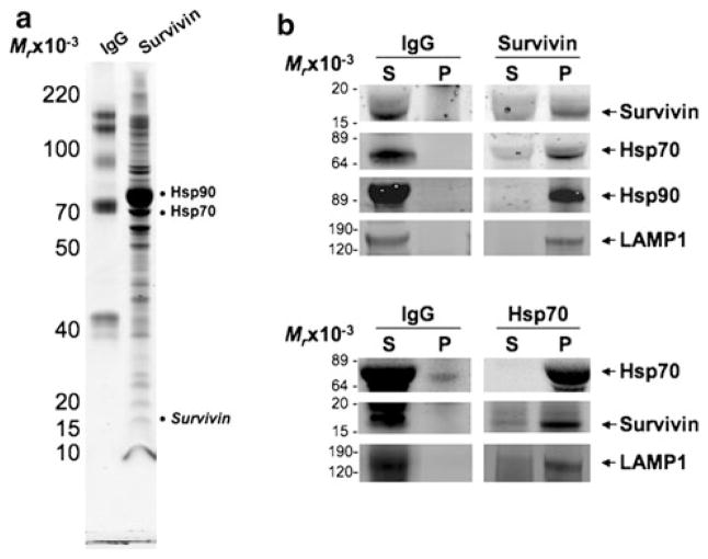
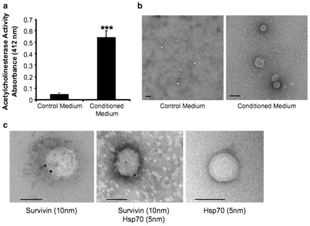
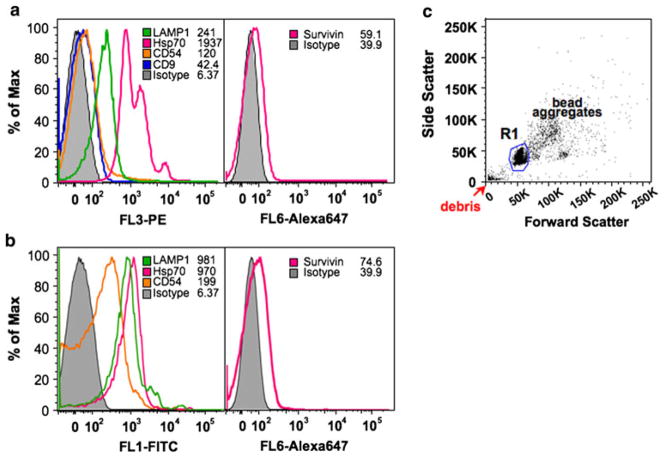
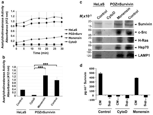
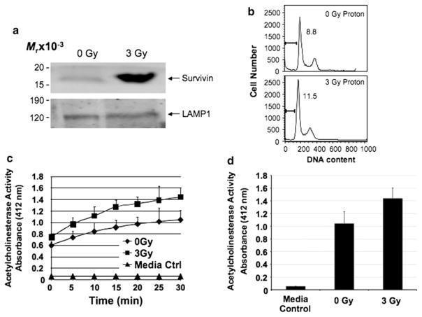
Inhibitor of apoptosis (IAP) and Heat shock proteins (HSPs) provide assistance in protecting cells from stresses of hypoxia, imbalanced pH, and altered metabolic and redox states commonly found in the microenvironmental mixture of tumor and nontumor cells. HSPs are upregulated, cell-surface displayed and released extracellularly in some types of tumors, a finding that until now was not shared by members of the IAP family. The IAP Survivin has been implicated in apoptosis inhibition and the regulation of mitosis in cancer cells. Survivin exists in a number of subcellular locations such as the mitochondria, cytoplasm, nucleus, and most recently, the extracellular space. Our previous work showing that extracellular survivin was able to enhance cellular proliferation, survival and tumor cell invasion provides evidence that Survivin might be secreted via an unidentified exocytotic pathway. In the present study, we describe for the first time the exosome-release of Survivin to the extracellular space both basally and after proton irradiation-induced stress. To examine whether exosomes contributed to Survivin release from cancer cells, exosomes were purified from HeLa cervical carcinoma cells and exosome quantity and Survivin content were determined. We demonstrate that although proton irradiation does not influence the exosomal secretory rate, the Survivin content of exosomes isolated from HeLa cells treated with a sublethal dose of proton irradiation (3 Gy) is significantly higher than control. These data identify a novel secretory pathway by which Survivin can be actively released from cells in both the basal and stress-induced state.


Conflict of interest statement

Conflict of interest The authors declare that they have no conflict of interest.

Figures

Fig. 1
Fig. 1
Survivin interacts with HSPs from conditioned medium. a Survivin and control IgG immunoprecipitates from HeLaS/POZn-Survivin conditioned medium were separated on a 4–12% gradient SDS gel and stained with Coomassie brilliant blue. The major bands were analyzed by trypsin digestion and LC–MS/MS mass spectrometry (see Supplemental Table 1). b Exosomes purified from the conditioned medium of HeLaS/POZnSurvivin cells were lysed and immunoprecipitated with pAb Survivin or pAb Hsp70 and pAb IgG. Aliquots of pellet (P) or supernatant (S) were sequentially immuno-blotted with pAb Survivin or pAb Hsp70 or mAb Hsp90 or mAb LAMP1. Molecular-weight markers in kilodaltons (KDa) are shown on the left
Fig. 2
Fig. 2
HeLaS/POZnSurvivin cells secrete Survivin and Hsp70-containing exosomes. a Exosome presence, measured as AChE activity from either 24 h control media or conditioned medium fractions. The 110,000g HeLaS/POZnSurvivin pellets were defined for AChE activity as described in the “Methods and materials”. Each fraction was evaluated by colorimetry. Values represent the means of three media control samples and nine conditioned medium samples. *** P <0.001. b Exosomes were characterized from 24 h HeLaS/POZnSurvivin conditioned medium using electron microscopy from both nonconditioned (left) and conditioned medium (right) respectively. c Immunoelectron microscopy on anti-Survivin- and anti-Hsp70-stained cryosections of HeLaS/POZnSurvivin cells shows that Survivin and Hsp70 localization in the exosome. Immunogold nanoparticles of 10 nm were conjugated to anti-Survivin pAb while 5 nm nanoparticles were conjugated to anti-Hsp70 mAb. Exosomes are cup-shaped and 50–150 nm in size. Bar 100 nm
Fig. 3
Fig. 3
Histogram profile of surface CD antigens on purified exosomes bound to anti-MHC class II (a) or CD9 (b) -coupled aldehyde beads. Presence of LAMP1, Hsp70, CD9, CD54 and Survivin antigens were analyzed using exosomes bound to anti-MHC class II- or CD9-coupled aldehyde beads. c Beads are identified on the forward and side scatter plot that represents single beads (R1), bead aggregates, and debris. The region R1 denotes the region selected for gating in all the experiments. Typically, single beads represent 50–80% of total beads
Fig. 4
Fig. 4
Survivin’s exosomal release from cancer cells is blocked by cytoD but not monensin. 24 h exosome release is quantitated from HeLaS cells as control or HeLaS/POZnSurvivin cells treated with vehicle, monensin or cytoD. a Exosome presence was measured as AChE activity. Direct modulation of actin polymerization with cytoD reduced exosome release measured for 30 min while inhibition of the common secretory pathway using monensin had no effect. b Measured at 30 min, this reduction in exosome presence after cytoD treatment was significant when compared to both monensin and control samples. AChE activity values represent the means of three samples. Exosomes were collected after 24 h of treatment. c Western blots of Survivin, c-Src, H-Ras, Hsp70 and the exosome specific marker protein LAMP1 in the conditioned medium of exosome isolations. C-Src, H-Ras and Hsp70 were found and are known exosome-associated proteins while LAMP1 is exosome specific. As in A, cytoD prohibited exosome proteins from accumulating in the exosomes while monensin did not. Protein concentrations for Western blot loading were determined using the AChE activity. Molecular-weight markers in kilodaltons (kDa) are shown on the left. *** P <0.001. d ELISA quantitation was accomplished according to the manufacturer’s instructions. Survivin was quantitated from conditioned medium (CM) and from CM having exosome removed using ultracentrifugation (Sup-supernatant) in the presence and absence of cytoD and monensin
Fig. 5
Fig. 5
Stress-activated Survivin protein content increases in exosomes after proton irradiation of HeLaS/POZnSurvivin cells. a Western blotting shows an increase in Survivin in exosome vesicles after 3 Gy of proton irradiation. Protein concentrations for Western blot loading were determined using the AChE activity. Loading is controlled for by the Lysosome Associated Membrane Protein (LAMP1). Molecular-weight markers in kilodaltons (KDa) are shown on the left. Exosomes were collected after 24 h of treatment. b Cervical carcinoma HeLaS/POZnSurvivin cells were irradiated using proton radiation (0 or 3 Gy) and after 24 h were harvested and analyzed for DNA content by propidium iodide staining and flow cytometry. Percentages of apoptotic cells with hypodiploid (sub-G1) DNA content or 4 N DNA representative of cells in G2/M are indicated per each condition tested. Data are representative of one of two independent experiments with comparable results. c Exosomes were purified from control media, 0 Gy and 3 Gy proton-irradiated HeLaS/POZnSurvivin conditioned medium after which AChE activity was determined as described under “Methods and materials”. Values represent the means of three samples. d Measured at 30 min, no significant difference was recorded

References

    1. Aznavoorian S, Stracke ML, Krutzsch H, Schiffmann E, Liotta LA. Signal transduction for chemotaxis and haptotaxis by matrix molecules in tumor cells. J Cell Biol. 1990;110:1427–1438. - PMC - PubMed
    1. Ohtani H. Stromal reaction in cancer tissue: pathophysiologic significance of the expression of matrix-degrading enzymes in relation to matrix turnover and immune/inflammatory reactions. Pathol Int. 1998;48:1–9. - PubMed
    1. Graner MW, Cumming RI, Bigner DD. The heat shock response and chaperones/heat shock proteins in brain tumors: surface expression, release, and possible immune consequences. J Neurosci. 2007;27:11214–11227. - PMC - PubMed
    1. Eustace BK, Jay DG. Extracellular roles for the molecular chaperone, hsp90. Cell Cycle. 2004;3:1098–1100. - PubMed
    1. Radons J, Multhoff G. Immunostimulatory functions of membrane-bound and exported heat shock protein 70. Exerc Immunol Rev. 2005;11:17–33. - PubMed

Publication types

MeSH terms