

Case Reports
doi: 10.1007/s11999-010-1528-9.
Orthopaedic case of the month: A 30-year-old woman with a painful forearm mass
Affiliations
- PMID: 20717855
- PMCID: PMC2947692
- DOI: 10.1007/s11999-010-1528-9
Item in Clipboard
Case Reports
Orthopaedic case of the month: A 30-year-old woman with a painful forearm mass
Clin Orthop Relat Res.
2010 Nov.
No abstract available
Figures

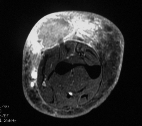
An axial T1-weighted MR image shows the somewhat poorly defined mass in the dorsal subcutaneous soft tissues of the forearm, which is isointense to low in signal compared with the underlying muscle. The high signal centrally likely attributable to hemorrhage and the prominent surrounding low signal edema can be seen.

An axial T2-weighted MR image with fat saturation shows the heterogeneous, but predominantly high signal mass with a thin, peripheral low-signal-intensity rim and marked surrounding soft tissue edema.
An axial T1-weighted MR image with fat saturation after the intravenous administration of gadolinium shows heterogeneous enhancement of the mass and exuberant enhancement in the surrounding soft tissues.

A photomicrograph shows frequent round to polygonal tumor cells with ample light cytoplasm and well-defined cell borders (Stain, hematoxylin and eosin; original magnification, ×60).

Conspicuous vascular invasion by the lesion is shown (Stain, hematoxylin and eosin; original magnification, ×4).

Extensive lymphatic permeation by the lesion is shown (Stain, hematoxylin and eosin; original magnification, ×20).
Similar articles
-
Primary epithelioid angiosarcoma of the lung presenting as pulmonary hemorrhage.Asian Cardiovasc Thorac Ann. 2006 Feb;14(1):69-71. doi: 10.1177/021849230601400118. Asian Cardiovasc Thorac Ann. 2006. PMID: 16432125
-
PEComa, a not so rare tumor that can cause clinical and pathologic diagnostic confusion: a case report.Conn Med. 2007 Aug;71(7):399-401. Conn Med. 2007. PMID: 17879861 No abstract available.
-
Juxtathyroidal neck soft tissue angiosarcoma presenting as an undifferentiated thyroid carcinoma.Thyroid. 2002 May;12(5):427-32. doi: 10.1089/105072502760043521. Thyroid. 2002. PMID: 12097205
-
Epithelioid angiosarcoma of the bladder: report of a new case with immunohistochemical profile and review of the literature.Pathology. 2011 Apr;43(3):290-3. doi: 10.1097/PAT.0b013e328344e2fb. Pathology. 2011. PMID: 21436648 Review. No abstract available.
-
Epithelioid malignant peripheral nerve sheath tumour: case report and review of the previously published cases.Cytopathology. 2002 Feb;13(1):54-63. doi: 10.1046/j.1365-2303.2002.00368.x. Cytopathology. 2002. PMID: 11985569 Review. No abstract available.
References
-
- Amo Y, Masuzawa M, Hamada Y, Katsuoka K. Serum concentrations of vascular endothelial growth factor-D in angiosarcoma patients. Br J Dermatol. 2004;150:160–161. - PubMed
-
- Breiteneder-Geleff S, Soleiman A, Kowalski H, Horvat R, Amann G, Kriehuber E, Diem K, Weninger W, Tschachler E, Alitalo K, Kerjaschki D. Angiosarcomas express mixed endothelial phenotypes of blood and lymphatic capillaries: podoplanin as a specific marker for lymphatic endothelium. Am J Pathol. 1999;154:385–394. - PMC - PubMed
-
- Budd GT. Management of angiosarcoma. Curr Oncol Rep. 2002;4:515–519. - PubMed
-
- Dancey AL, Mahon BS, Rayatt SS. A review of diagnostic imaging in melanoma. J Plast Reconstr Aesthet Surg. 2008;61:1275–1283. - PubMed
-
- DeMartelaere SL, Roberts D, Burgess MA, Morrison WH, Pisters PW, Sturgis EM, Ho V, Esmaeli B. Neoadjuvant chemotherapy-specific and overall treatment outcomes in patients with cutaneous angiosarcoma of the face with periorbital involvement. Head Neck. 2008;30:639–646. - PubMed
Publication types
MeSH terms
LinkOut - more resources
Full Text Sources
Medical
