Combined technologies for microfabricating elastomeric cardiac tissue engineering scaffolds
- PMID: 20718054
- PMCID: PMC3315382
- DOI: 10.1002/mabi.201000165
Combined technologies for microfabricating elastomeric cardiac tissue engineering scaffolds
Abstract
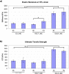
Polymer scaffolds that direct elongation and orientation of cultured cells can enable tissue engineered muscle to act as a mechanically functional unit. We combined micromolding and microablation technologies to create muscle tissue engineering scaffolds from the biodegradable elastomer poly(glycerol sebacate). These scaffolds exhibited well defined surface patterns and pores and robust elastomeric tensile mechanical properties. Cultured C2C12 muscle cells penetrated the pores to form spatially controlled engineered tissues. Scanning electron and confocal microscopy revealed muscle cell orientation in a preferential direction, parallel to micromolded gratings and long axes of microablated anisotropic pores, with significant individual and interactive effects of gratings and pore design.
Figures


Similar articles
-
Laser microfabricated poly(glycerol sebacate) scaffolds for heart valve tissue engineering.J Biomed Mater Res A. 2013 Jan;101(1):104-14. doi: 10.1002/jbm.a.34305. Epub 2012 Jul 24. J Biomed Mater Res A. 2013. PMID: 22826211 Free PMC article.
-
3D structural patterns in scalable, elastomeric scaffolds guide engineered tissue architecture.Adv Mater. 2013 Aug 27;25(32):4459-65. doi: 10.1002/adma.201301016. Epub 2013 Jun 14. Adv Mater. 2013. PMID: 23765688 Free PMC article.
-
Highly elastomeric poly(glycerol sebacate)-co-poly(ethylene glycol) amphiphilic block copolymers.Biomaterials. 2013 May;34(16):3970-3983. doi: 10.1016/j.biomaterials.2013.01.045. Epub 2013 Mar 1. Biomaterials. 2013. PMID: 23453201 Free PMC article.
-
Valvular interstitial cell seeded poly(glycerol sebacate) scaffolds: toward a biomimetic in vitro model for heart valve tissue engineering.Acta Biomater. 2013 Apr;9(4):5974-88. doi: 10.1016/j.actbio.2013.01.001. Epub 2013 Jan 5. Acta Biomater. 2013. PMID: 23295404
-
Preparation of aligned poly(glycerol sebacate) fibrous membranes for anisotropic tissue engineering.Mater Sci Eng C Mater Biol Appl. 2019 Jul;100:30-37. doi: 10.1016/j.msec.2019.02.098. Epub 2019 Feb 27. Mater Sci Eng C Mater Biol Appl. 2019. PMID: 30948065
Cited by
-
Mechanical properties of murine and porcine ocular tissues in compression.Exp Eye Res. 2014 Apr;121:194-9. doi: 10.1016/j.exer.2014.02.020. Epub 2014 Mar 5. Exp Eye Res. 2014. PMID: 24613781 Free PMC article.
-
A biodegradable microvessel scaffold as a framework to enable vascular support of engineered tissues.Biomaterials. 2013 Dec;34(38):10007-15. doi: 10.1016/j.biomaterials.2013.09.039. Epub 2013 Sep 27. Biomaterials. 2013. PMID: 24079890 Free PMC article.
-
Biomimetic scaffold combined with electrical stimulation and growth factor promotes tissue engineered cardiac development.Exp Cell Res. 2014 Feb 15;321(2):297-306. doi: 10.1016/j.yexcr.2013.11.005. Epub 2013 Nov 14. Exp Cell Res. 2014. PMID: 24240126 Free PMC article.
-
Laser microfabricated poly(glycerol sebacate) scaffolds for heart valve tissue engineering.J Biomed Mater Res A. 2013 Jan;101(1):104-14. doi: 10.1002/jbm.a.34305. Epub 2012 Jul 24. J Biomed Mater Res A. 2013. PMID: 22826211 Free PMC article.
-
The significance of pore microarchitecture in a multi-layered elastomeric scaffold for contractile cardiac muscle constructs.Biomaterials. 2011 Mar;32(7):1856-64. doi: 10.1016/j.biomaterials.2010.11.032. Epub 2010 Dec 8. Biomaterials. 2011. PMID: 21144580 Free PMC article.
References
Publication types
MeSH terms
Substances
Grants and funding
LinkOut - more resources
Full Text Sources

