

Autonomous in vitro anticancer drug release from mesoporous silica nanoparticles by pH-sensitive nanovalves
- PMID: 20718462
- PMCID: PMC3116646
- DOI: 10.1021/ja104501a
Autonomous in vitro anticancer drug release from mesoporous silica nanoparticles by pH-sensitive nanovalves
Abstract
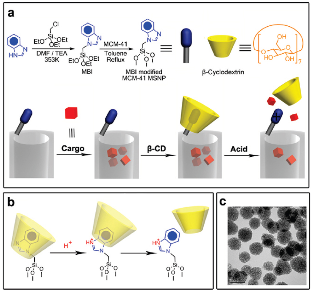
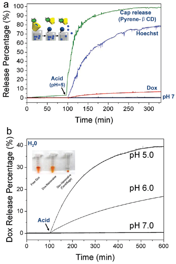
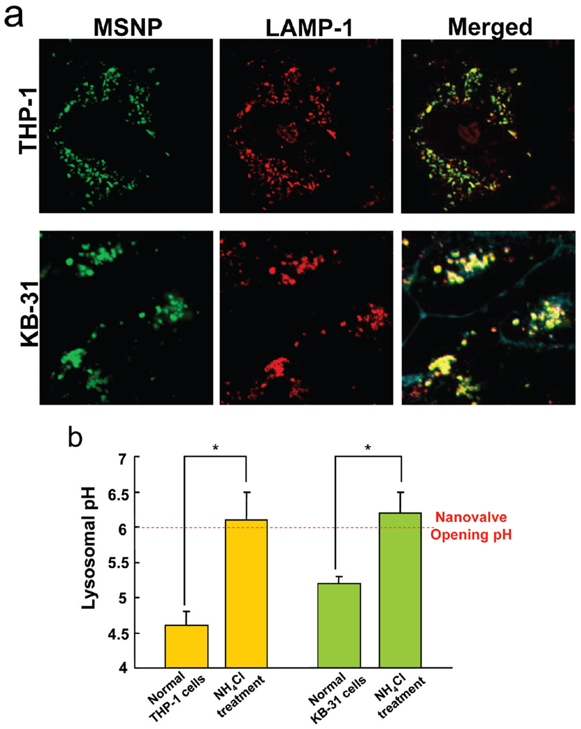
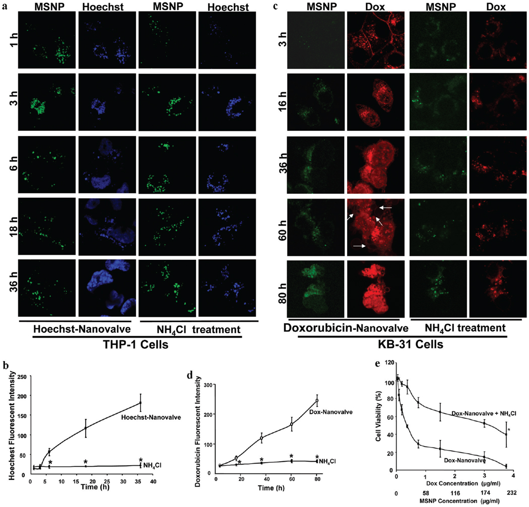
Mesoporous silica nanoparticles (MSNP) have proven to be an extremely effective solid support for controlled drug delivery on account of the fact that their surfaces can be easily functionalized in order to control the nanopore openings. We have described recently a series of mechanized silica nanoparticles, which, under abiotic conditions, are capable of delivering cargo molecules employing a series of nanovalves. The key question for these systems has now become whether they can be adapted for biological use through controlled nanovalve opening in cells. Herein, we report a novel MSNP delivery system capable of drug delivery based on the function of beta-cyclodextrin (beta-CD) nanovalves that are responsive to the endosomal acidification conditions in human differentiated myeloid (THP-1) and squamous carcinoma (KB-31) cell lines. Furthermore, we demonstrate how to optimize the surface functionalization of the MSNP so as to provide a platform for the effective and rapid doxorubicin release to the nuclei of KB-31 cells.
Figures
Similar articles
-
Functional nanovalves on protein-coated nanoparticles for in vitro and in vivo controlled drug delivery.Small. 2015 Jan 21;11(3):319-328. doi: 10.1002/smll.201400765. Epub 2014 Sep 5. Small. 2015. PMID: 25196485 Free PMC article.
-
A dual-functional HER2 aptamer-conjugated, pH-activated mesoporous silica nanocarrier-based drug delivery system provides in vitro synergistic cytotoxicity in HER2-positive breast cancer cells.Int J Nanomedicine. 2019 May 31;14:4029-4044. doi: 10.2147/IJN.S201688. eCollection 2019. Int J Nanomedicine. 2019. PMID: 31213813 Free PMC article.
-
Dual stimuli-responsive nano-vehicles for controlled drug delivery: mesoporous silica nanoparticles end-capped with natural chitosan.Chem Commun (Camb). 2014 Nov 11;50(87):13268-71. doi: 10.1039/c4cc04383a. Epub 2014 Sep 19. Chem Commun (Camb). 2014. PMID: 25237679
-
Tailored Mesoporous Silica Nanoparticles for Controlled Drug Delivery: Platform Fabrication, Targeted Delivery, and Computational Design and Analysis.Mini Rev Med Chem. 2018;18(11):976-989. doi: 10.2174/1389557516666160505114814. Mini Rev Med Chem. 2018. PMID: 27145854 Review.
-
Gated Materials: Installing Macrocyclic Arenes-Based Supramolecular Nanovalves on Porous Nanomaterials for Controlled Cargo Release.Biotechnol J. 2019 Jan;14(1):e1800354. doi: 10.1002/biot.201800354. Epub 2018 Nov 26. Biotechnol J. 2019. PMID: 30457707 Review.
Cited by
-
Multifunctional hybrid silica nanoparticles for controlled doxorubicin loading and release with thermal and pH dually response.J Mater Chem B. 2013;1(8):1109-1118. doi: 10.1039/C2TB00223J. Epub 2012 Dec 13. J Mater Chem B. 2013. PMID: 23543911 Free PMC article.
-
Combination delivery of TGF-β inhibitor and IL-2 by nanoscale liposomal polymeric gels enhances tumour immunotherapy.Nat Mater. 2012 Oct;11(10):895-905. doi: 10.1038/nmat3355. Epub 2012 Jul 15. Nat Mater. 2012. PMID: 22797827 Free PMC article.
-
Functioning of nanovalves on polymer coated mesoporous silica Nanoparticles.Nanoscale. 2013 Nov 7;5(21):10300-6. doi: 10.1039/c3nr03442a. Epub 2013 Sep 9. Nanoscale. 2013. PMID: 24056925 Free PMC article.
-
Exosome encased spherical nucleic acid gold nanoparticle conjugates as potent microRNA regulation agents.Small. 2014 Jan 15;10(1):186-92. doi: 10.1002/smll.201302143. Epub 2013 Sep 17. Small. 2014. PMID: 24106176 Free PMC article.
-
Nanovalve activation by surface-attached photoacids.Chem Commun (Camb). 2014 Aug 7;50(61):8388-90. doi: 10.1039/c4cc03293d. Chem Commun (Camb). 2014. PMID: 24942753 Free PMC article.
References
-
- Kresge CT, Leonowicz ME, Roth WJ, Vartuli JC, Beck JS. Nature. 1992;359:710–712.
- Cai Q, Luo ZS, Pang WQ, Fan YW, Chen XH, Cui FZ. Chem. Mater. 2001;13:258–263.
- Lai C-Y, Trewyn BG, Jeftinija DM, Jeftinija K, Xu S, Jeftinija S, Lin VSY. J. Am. Chem. Soc. 2003;125:4451–4459. - PubMed
-
- Cotí KK, Belowich ME, Liong M, Ambrogio MW, Lau YA, Khatib HA, Zink JI, Khashab NM, Stoddart JF. Nanoscale. 2009;1:16–39. - PubMed
- Trewyn BG, Slowing II, Giri S, Chen HT, Lin VSY. Acc. Chem. Res. 2007;40:846–853. - PubMed
- Slowing II, Trewyn BG, Giri S, Lin VS-Y. Adv. Funct. Mater. 2007;17:1225–1236.
- Slowing I, Vivero-Escoto J, Wu C-W, Lin V. Adv. Drug Delivery Rev. 2008;60:1278–1288. - PubMed
-
- Zhu YF, Shi JL, Shen WH, Dong XP, Feng JW, Ruan ML, Li YS. Angew. Chem., Int. Ed. 2005;44:5083–5087. - PubMed
- Zhu C-L, Song X-Y, Zhou W-H, Yang H-H, Wen Y-H, Wang X-R. J. Mater. Chem. 2009;19:7765–7770.
- Andrew MacKay J, Chen M, McDaniel JR, Liu W, Simnick AJ, Chilkoti A. Nat. Mater. 2009;8:993–999. - PMC - PubMed
-
- Lu J, Choi E, Tamanoi F, Zink JI. Small. 2008;4:421–-426. - PMC - PubMed
- Patel K, Angelos S, Dichtel WR, Coskun A, Yang YW, Zink JI, Stoddart JF. J. Am. Chem. Soc. 2008;130:2382–2383. - PubMed
- Nguyen TD, Leung KCF, Liong M, Pentecost CD, Stoddart JF, Zink JI. Org. Lett. 2006;8:3363–3366. - PubMed
- Angelos S, Yang YW, Patel K, Stoddart JF, Zink JI. Angew. Chem., Int. Ed. 2008;47:2222–2226. - PubMed
- Du L, Liao S, Khatib HA, Stoddart JF, Zink JI. J. Am. Chem. Soc. 2009;131:15136–15142. - PubMed
- Angelos S, Khashab NM, Yang YW, Trabolsi A, Khatib HA, Stoddart JF, Zink JI. J. Am. Chem. Soc. 2009;131:12912–12914. - PubMed
- Liu R, Zhang Y, Zhao X, Agarwal A, Mueller LJ, Feng P. J. Am. Chem. Soc. 2010;132:1500–1501. - PubMed
Publication types
MeSH terms
Substances
Grants and funding
LinkOut - more resources
Full Text Sources
Other Literature Sources
