

Interaction of silver nanoparticles with Tacaribe virus
- PMID: 20718972
- PMCID: PMC2936366
- DOI: 10.1186/1477-3155-8-19
Interaction of silver nanoparticles with Tacaribe virus
Abstract
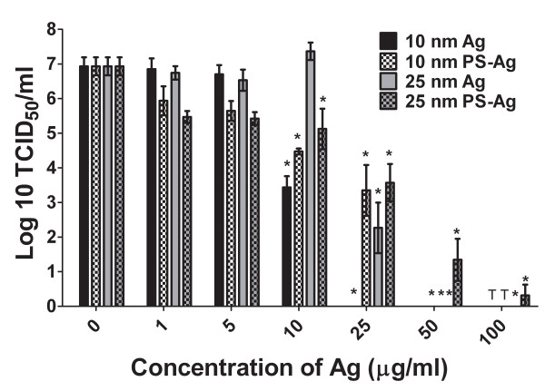
Background: Silver nanoparticles possess many unique properties that make them attractive for use in biological applications. Recently they received attention when it was shown that 10 nm silver nanoparticles were bactericidal, which is promising in light of the growing number of antibiotic resistant bacteria. An area that has been largely unexplored is the interaction of nanomaterials with viruses and the possible use of silver nanoparticles as an antiviral agent.
Results: This research focuses on evaluating the interaction of silver nanoparticles with a New World arenavirus, Tacaribe virus, to determine if they influence viral replication. Surprisingly exposing the virus to silver nanoparticles prior to infection actually facilitated virus uptake into the host cells, but the silver-treated virus had a significant reduction in viral RNA production and progeny virus release, which indicates that silver nanoparticles are capable of inhibiting arenavirus infection in vitro. The inhibition of viral replication must occur during early replication since although pre-infection treatment with silver nanoparticles is very effective, the post-infection addition of silver nanoparticles is only effective if administered within the first 2-4 hours of virus replication.
Conclusions: Silver nanoparticles are capable of inhibiting a prototype arenavirus at non-toxic concentrations and effectively inhibit arenavirus replication when administered prior to viral infection or early after initial virus exposure. This suggests that the mode of action of viral neutralization by silver nanoparticles occurs during the early phases of viral replication.
Figures


Similar articles
-
Inhibition effect of silver nanoparticles on herpes simplex virus 2.Genet Mol Res. 2014 Mar 19;13(3):7022-8. doi: 10.4238/2014.March.19.2. Genet Mol Res. 2014. PMID: 24682984
-
Silver nanoparticles inhibit hepatitis B virus replication.Antivir Ther. 2008;13(2):253-62. Antivir Ther. 2008. PMID: 18505176
-
Silver nanoparticles inhibit vaccinia virus infection by preventing viral entry through a macropinocytosis-dependent mechanism.J Biomed Nanotechnol. 2013 Sep;9(9):1624-35. doi: 10.1166/jbn.2013.1659. J Biomed Nanotechnol. 2013. PMID: 23980510
-
Silver Nanoparticles as Potential Antiviral Agents.Pharmaceutics. 2021 Nov 29;13(12):2034. doi: 10.3390/pharmaceutics13122034. Pharmaceutics. 2021. PMID: 34959320 Free PMC article. Review.
-
Metal nanoparticles: The protective nanoshield against virus infection.Crit Rev Microbiol. 2016;42(1):46-56. doi: 10.3109/1040841X.2013.879849. Epub 2014 Apr 22. Crit Rev Microbiol. 2016. PMID: 24754250 Review.
Cited by
-
Fighting arboviral diseases: low toxicity on mammalian cells, dengue growth inhibition (in vitro), and mosquitocidal activity of Centroceras clavulatum-synthesized silver nanoparticles.Parasitol Res. 2016 Feb;115(2):651-62. doi: 10.1007/s00436-015-4783-6. Parasitol Res. 2016. PMID: 26462804
-
Antiviral and Immunomodulatory Activity of Silver Nanoparticles in Experimental RSV Infection.Viruses. 2019 Aug 8;11(8):732. doi: 10.3390/v11080732. Viruses. 2019. PMID: 31398832 Free PMC article.
-
Protective hybrid coating containing silver, copper and zinc cations effective against human immunodeficiency virus and other enveloped viruses.BMC Microbiol. 2016 Apr 1;16 Suppl 1:56. doi: 10.1186/s12866-016-0675-x. BMC Microbiol. 2016. PMID: 27036553 Free PMC article.
-
Antiviral Activity of Graphene Oxide-Silver Nanocomposites Against Murine Betacoronavirus.Int J Nanomedicine. 2024 Sep 4;19:9009-9033. doi: 10.2147/IJN.S473448. eCollection 2024. Int J Nanomedicine. 2024. PMID: 39246425 Free PMC article.
-
Silver nanoparticles: synthesis, characterisation and biomedical applications.Open Life Sci. 2020 Nov 19;15(1):819-839. doi: 10.1515/biol-2020-0094. eCollection 2020. Open Life Sci. 2020. PMID: 33817269 Free PMC article. Review.
References
-
- Pedras-Vasconcelos JA, Goucher D, Puig M, Tonelli LH, Wang V, Ito S, Verthelyi D. CpG Oligodeoxynucleotides Protect Newborn Mice from a Lethal Challenge with the Neurotropic Tacaribe Arenavirus. J Immunol. 2006;176:4940–4949. - PubMed
LinkOut - more resources
Full Text Sources
