. 2010;19(12):1645-57.
doi: 10.3727/096368910X516628. Epub 2010 Aug 17.

Delivery of a therapeutic protein by immune-privileged Sertoli cells

Affiliations

Delivery of a therapeutic protein by immune-privileged Sertoli cells

Katelyn Halley et al. Cell Transplant. 2010.

Abstract

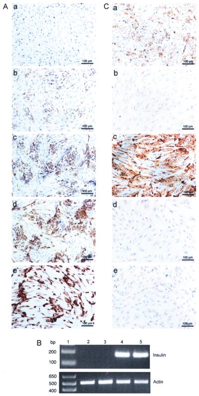
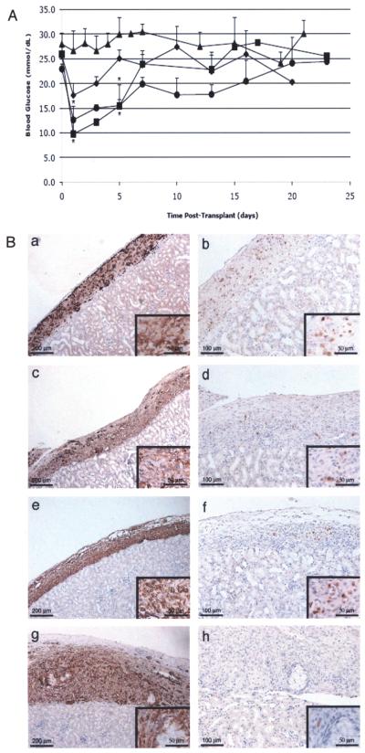
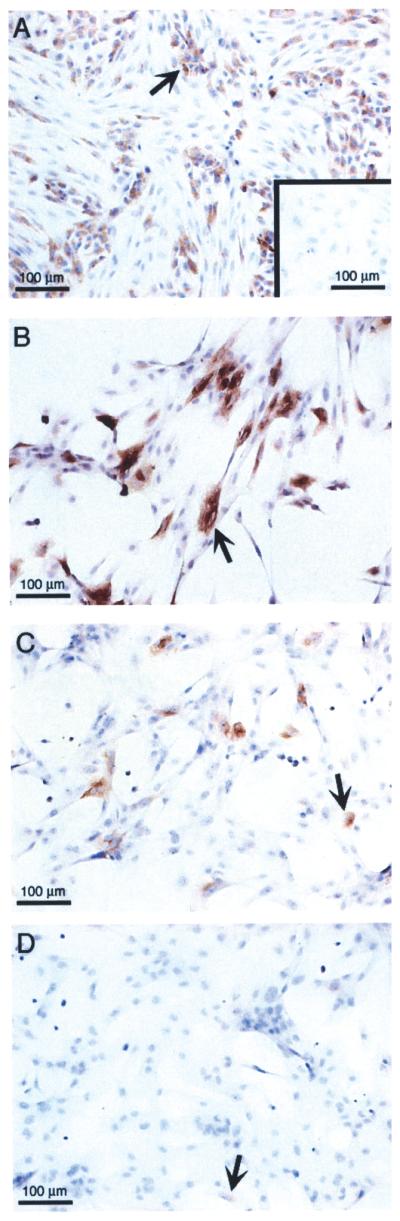
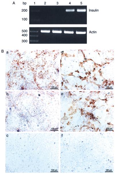
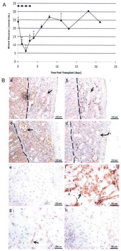
Immune-privileged Sertoli cells survive long term after allogeneic or xenogeneic transplantation without the use of immunosuppressive drugs, suggesting they could be used as a vehicle to deliver therapeutic proteins. As a model to test this, we engineered Sertoli cells to transiently produce basal levels of insulin and then examined their ability to lower blood glucose levels after transplantation into diabetic SCID mice. Mouse and porcine Sertoli cells transduced with a recombinant adenoviral vector containing furin-modified human proinsulin cDNA expressed insulin mRNA and secreted insulin protein. Transplantation of 5-20 million insulin-expressing porcine Sertoli cells into diabetic SCID mice significantly decreased blood glucose levels in a dose-dependent manner, with 20 million Sertoli cells decreasing blood glucose levels to 9.8 ± 2.7 mM. Similar results were obtained when 20 million insulin-positive, BALB/c mouse Sertoli cells were transplanted; blood glucose levels dropped to 6.3 ± 2.4 mM and remained significantly lower for 5 days. To our knowledge, this is the first study to demonstrate Sertoli cells can be engineered to produce and secrete a clinically relevant factor that has a therapeutic effect, thus supporting the concept of using immune-privileged Sertoli cells as a potential vehicle for gene therapy.


Figures

Figure 1
Figure 1
Production of insulin by adenovirus transduced neonatal porcine Sertoli cells. (A, C) NPSC were cultured overnight as a monolayer on chamber slides, transduced with AdCMVhInsM at a MOI of 0 (A, a), 50 (A, b), 75 (A, c), 100 (A, d and C, a), or 200 (A, e), AdCMVhInsWT (C, b and c) at a MOI of 100 or AdRSVGFP (C, d and e) at a MOI of 100. Slides were collected after 2 days, fixed with 1% paraformaldehyde, and immunostained for insulin (A, a–e and C, b and d) or C-peptide (C, a, c, and e). All sections were counterstained with hematoxylin. (B) NPSC cultured overnight as a monolayer on six-well plates with DMEM plus 10% FBS were either nontransduced (MOI of 0; lane 2) or transduced with AdRSVGFP (lane 3), AdCMVhInsWT (lane 4), or AdCMVhInsM (lane 5) adenoviral vectors at a MOI of 100. Cells were collected after 2 days and RT-PCR was performed for insulin or β-actin. Lane 1 is the 1 kb Plus DNA Ladder (Invitrogen).
Figure 2
Figure 2
Transplantation of insulin-expressing neonatal porcine Sertoli cells into diabetic SCID mice. (A) Average blood glucose values ± SEM of SCID mice after transplantation with 5 (filled diamonds; n = 5), 10 (filled circles; n = 4), or 20 (filled squares; n = 3) million insulin-expressing NPSC or 20 million (filled triangles; n = 4) GFP-expressing NPSC. NPSC were transduced with the AdCMVhInsM or AdRSVGFP adenoviral vector at a MOI of 100. *Mean blood glucose values were significantly decreased compared to the AdRSVGFP group values (p < 0.05). (B) Grafts were removed from SCID mice at 3 (a and b), 8 (c and d), 13 (e and f), and 30 (g and h) days after transplantation with NPSC and immunostained for the SC marker vimentin (column 1; a, c, e, and g) or insulin (column 2; b, d, f, and h). Higher magnification photomicrographs are included as insets (a–h). All sections were counterstained with hematoxylin.
Figure 3
Figure 3
Long-term culture of neonatal porcine Sertoli cells transduced with the furin-modified adenoviral vector. NPSC were cultured overnight as a monolayer on chamber slides, transduced with AdCMVhInsM at a MOI of 0 (A, inset) or 100 (A–D) and collected after 5 (A), 12 (B), 15 (C), or 17 (D) days. Slides were fixed with 1% paraformaldehyde and immunostained for insulin. All sections were counterstained with hematoxylin. Arrow shows insulin-positive SC.
Figure 4
Figure 4
In vitro expression of insulin and C-peptide or proinsulin by adenovirus transduced mouse Sertoli cells. (A) mSC cultured overnight as a monolayer on six-well plates with DMEM plus 10% FBS were either nontransduced (MOI of 0; lane 2) or transduced with AdRSVGFP (lane 3), AdCMVhInsWT (lane 4), or AdCMVhInsM (lane 5) adenoviral vectors at a MOI of 100. Cells were collected after 2 days and RT-PCR was performed for insulin or β-actin. Lane 1 is the 1kb Plus DNA Ladder (Invitrogen). (B) mSC cultured overnight as a monolayer on chamber slides were transduced with AdCMVhInsM (a and d), AdCMVhInsWT (b and e), or AdRSVGFP (c and f) adenoviral vectors at a MOI of 100. Slides were collected after 2 days, fixed with 1% paraformaldehyde and immunostained for insulin (a–c) or C-peptide (d–f). All sections were counterstained with hematoxylin.
Figure 5
Figure 5
Transplantation of insulin-expressing mouse Sertoli cells and immunohistochemical analysis of Sertoli cells transduced with the furin-modified adenoviral vector. (A) Average blood glucose values ± SEM of SCID mice after transplantation with 20 million mSC. mSC were transduced with AdCMVhInsM (filled diamonds; n = 3) or AdRSVGFP (filled squares; n = 1) at a MOI of 100. *Mean blood glucose values were significantly decreased compared to pretransplant values (p < 0.05). (B) mSC grafts were removed from SCID mice at 8 (a and b) and 30 (c and d) days after transplantation with mSC and immunostained for the SC marker GATA-4 (a and c) or insulin (b and d). The graft and kidney are separated by a dotted line. mSC cultured overnight as a monolayer on chamber slides were transduced with AdCMVhInsM at a MOI of 0 (e) or 100 (f–h) and collected after 5 (e and f), 12 (g), or 20 (h) days. Slides were fixed with 1% paraformaldehyde and immunostained for insulin (e-h). All sections were counterstained with hematoxylin. Arrow shows SC.

Similar articles

Cited by

References

    1. Barry SC, Ramesh N, Lejnieks D, Simonson WT, Kemper L, Lernmark A, Osborne WR. Glucose-regulated insulin expression in diabetic rats. Hum. Gene Ther. 2001;12(2):131–139. - PubMed
    1. Bellgrau D, Gold D, Selawry H, Moore J, Franzusoff A, Duke RC. A role for CD95 ligand in preventing graft rejection. Nature. 1995;377(6550):630–632. - PubMed
    1. Davidson HW. (Pro)Insulin processing: A historical perspective. Cell Biochem. Biophys. 2004;40(3 Suppl.):143–158. - PubMed
    1. Dong H, Altomonte J, Morral N, Meseck M, Thung SN, Woo SL. Basal insulin gene expression significantly improves conventional insulin therapy in type 1 diabetic rats. Diabetes. 2002;51(1):130–138. - PubMed
    1. Dufour JM, Dass B, Halley KR, Korbutt GS, Dixon DE, Rajotte RV. Sertoli cell line lacks the immunoprotective properties associated with primary Sertoli cells. Cell Transplant. 2008;17(5):525–534. - PubMed

Publication types

MeSH terms