. 2010 Sep 17;400(2):212-8.
doi: 10.1016/j.bbrc.2010.08.040. Epub 2010 Aug 16.

Human CD133-derived bone marrow stromal cells establish ectopic hematopoietic microenvironments in immunodeficient mice

Affiliations

Human CD133-derived bone marrow stromal cells establish ectopic hematopoietic microenvironments in immunodeficient mice

Benjamin Bakondi et al. Biochem Biophys Res Commun. .

Abstract

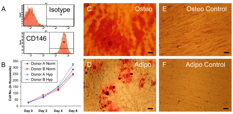
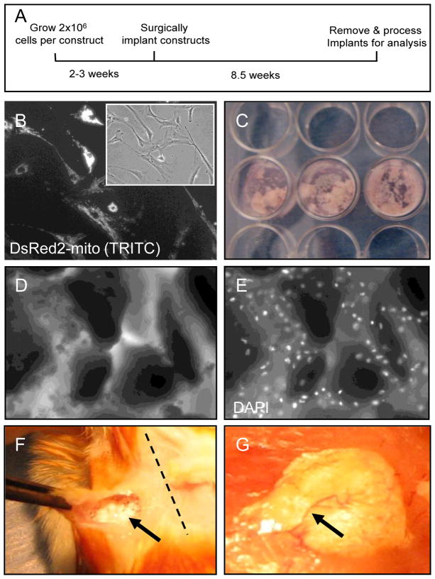
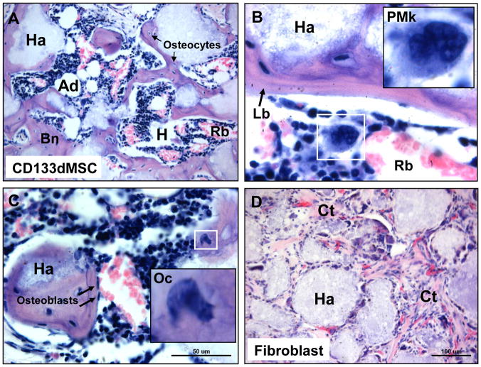
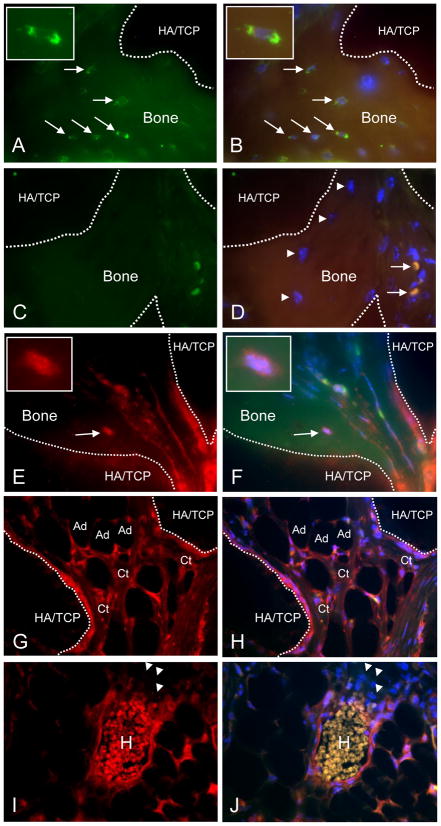
Cultured adherent bone marrow stromal cells (BMSCs) are capable of forming ectopic hematopoietic microenvironments (HMEs) in immunodeficient mice. However, the cell surface phenotype of the native bone marrow stem/progenitor cell that gives rise to BMSCs that support hematopoiesis remains poorly defined. We recently reported the derivation of human BMSC-like cells (CD133BMSCs) by magnetic cell sorting against Prominin-1 (CD133), an epitope expressed by embryonic, fetal, and adult stem cells. Here we demonstrate that CD133BMSCs are capable of forming ectopic HMEs. Cultured adherent CD133BMSCs derived from sorted CD133-positive cells lacked CD133 expression, but were uniformly positive for CD146, an epitope recently described to identify self-renewing osteoprogenitor cells that could transfer the HME. CD133BMSCs were genetically-tagged by lentivirus, expanded, and seeded into HA/TCP/fibrin constructs that were implanted subcutaneously. After 60days, CD133BMSCs produced human osteocytes, osteoblasts, adipocytes, and reticular cells that supported murine hematopoiesis. CD133BMSCs that were not transduced with lentivirus also formed HMEs. Control constructs seeded with human dermal fibroblasts formed connective tissue, but failed to form HMEs. Our data indicate that CD133 expression identifies a native human bone marrow stem/progenitor cell that gives rise to BMSCs capable of forming the HME.


Figures

Fig. 1
Fig. 1. Characterization of CD133BMSCs
A) Cell surface phenotyping for CD146 (MCAM). Note that isotype control is negative. B) High density growth of CD133BMSCs (N=2 donors) seeded at 1,042 cm2 in normoxic (21% oxygen; red lines) or hypoxic (1% oxygen; blue lines) conditions for 8 days. C–F) Phase contrast photomicrographs of cultured CD133BMSCs differentiated over 3 weeks into osteoblasts (Osteo) and adipocytes (Adipo). Calcium deposition was stained by Alizarin Red S (C) and lipid deposition was stained by Oil Red O (D). Alizarin Red S and Oil Red O staining was absent in CD133BMSCs treated with control growth medium (E, F). CF, 40X magnification.
Fig. 2
Fig. 2. Preparation and implantation of cell-seeded HA/TCP/fibrin constructs
A) Schematic of HME transfer protocol. B) Epifluorescent imaging of CD133BMSCs expressing DsRed2-mito reporter (20X magnification). Inset: phase contrast photomicrograph of CD133dMSCs pictured in (B). C) Picture of constructs prepared in 24-well plates and incubated for 90 minutes at 37ºC in a cell culture incubator prior to implantation. D) Phase contrast photomicrograph of HA/TCP particles incubated with cells and clotting factors (10X magnification). E) Epifluorescent image of (D). Note that CD133BMSCs are evenly dispersed throughout the construct. White: DAPI nuclear stain. F) At the time of surgery, gelatinous construct material was implanted into subcutaneous pockets (arrow) in immunodeficient mice at the scapula proximal or medial to the dorsal midline (dotted line). G) At 60 days after implantation, solidified constructs were integrated into host tissue and were vascularized (arrow).
Fig. 3
Fig. 3. Histology of cell-seeded constructs 60 days after implantation
A) H & E stain of CD133BMSC-seeded construct showing HME formation with blood forming hematopoietic cells (Hp), enucleated red blood cells (Rb), and adipocytes (Ad), that are interspersed throughout bone (Bn) (10X magnification). Note that bone surrounds the HA/TCP carrier (Ha). Osteocytes (Os) were observed residing within lacunae (arrows). B) Concentrically-layered lamellar bone (Lb, arrow) had formed on HA/TCP surfaces (Ha) (40X magnification). Inset: promegakaryocyte (PMk). C) Erythropoiesis at various stages was observed throughout the HMEs. Inset: osteoclast (Oc). Arrows: osteoblasts. D) HA/TCP/fibrin construct seeded with 2 × 10⁶ human fibroblasts. Fibroblasts adhered to the HA/TCP matrix (Ha) formed connective tissue (Ct) containing microvasculature throughout the implant, but did not support hematopoiesis.
Fig. 4
Fig. 4. Immunohistochemical evaluation of CD133dMSC-seeded constructs
A, B) Human-specific β2M staining (green, FITC channel) of osteocytes within bone formed from CD133BMSCs (arrows) (100X magnification). Dotted lines delineate bone (autofluorescent) and HA/TCP (dark areas). FITC/TRITC merge with DAPI (B). Insets: magnified view of an osteocyte. C, D) Isotype control staining (1 μg/ml IgG, 100X magnification). FITC/TRITC merge with DAPI (D). Arrowheads: osteocytes negative for staining by isotype. Arrows: strongly autofluorescent red blood cells. E, F) Staining for DsRed demonstrates characteristic punctate pattern of mitochondria within a bone-residing osteocyte (TRITC channel, E) and merge (F). Insets: DsRed-positive osteocyte. G, H) DsRed-positive inter-sinusoidal reticular cells, connective tissue (Ct), and adipocytes (Ad) in the implant. I, J) DsRed-positive peri-vascular reticular cells surrounding a sinus. H, Autofluorescent host-derived red blood cells. Arrowheads: Host-derived erythroblasts did not stain positive for DsRed. TRITC channel (E–I). TRITC/FITC merge with DAPI (F–J).

References

    1. Wilson A, Trumpp A. Bone-marrow haematopoietic-stem-cell niches. Nat Rev Immunol. 2006;6:93–106. - PubMed
    1. Zhang J, Niu C, Ye L, et al. Identification of the haematopoietic stem cell niche and control of the niche size. Nature. 2003;425:836–41. - PubMed
    1. Kiel MJ, Yilmaz OH, Iwashita T, et al. SLAM family receptors distinguish hematopoietic stem and progenitor cells and reveal endothelial niches for stem cells. Cell. 2005;121:1109–21. - PubMed
    1. Calvi LM, Adams GB, Weibrecht KW, et al. Osteoblastic cells regulate the haematopoietic stem cell niche. Nature. 2003;425:841–6. - PubMed
    1. Scadden DT. The stem-cell niche as an entity of action. Nature. 2006;441:1075–9. - PubMed

Publication types