

Acute myocardial rescue with endogenous endothelial progenitor cell therapy
- PMID: 20719564
- PMCID: PMC3235678
- DOI: 10.1016/j.hlc.2010.06.1056
Acute myocardial rescue with endogenous endothelial progenitor cell therapy
Abstract
Purpose: Post-myocardial infarction heart failure is a major health concern with limited therapy. Molecular revascularisation utilising granulocyte-macrophage colony stimulating factor (GMCSF) mediated endothelial progenitor cell (EPC) upregulation and stromal cell derived factor-1α (SDF) mediated myocardial EPC chemokinesis, may prevent myocardial loss and adverse remodelling. Vasculogenesis, viability, and haemodynamic improvements following therapy were investigated.
Procedures: Lewis rats (n=91) underwent LAD ligation and received either intramyocardial SDF and subcutaneous GMCSF or saline injections at the time of infarction. Molecular and haemodynamic assessments were performed at pre-determined time points following ligation.
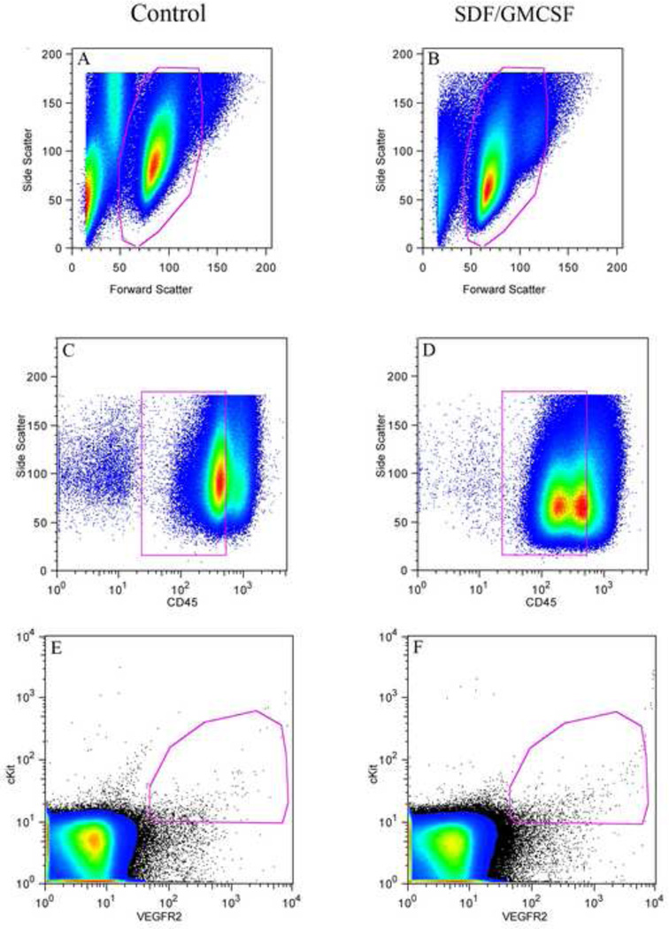
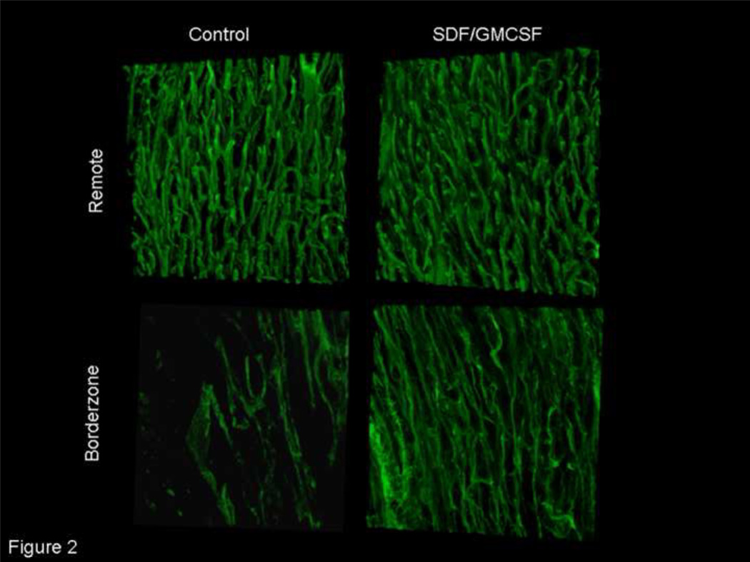
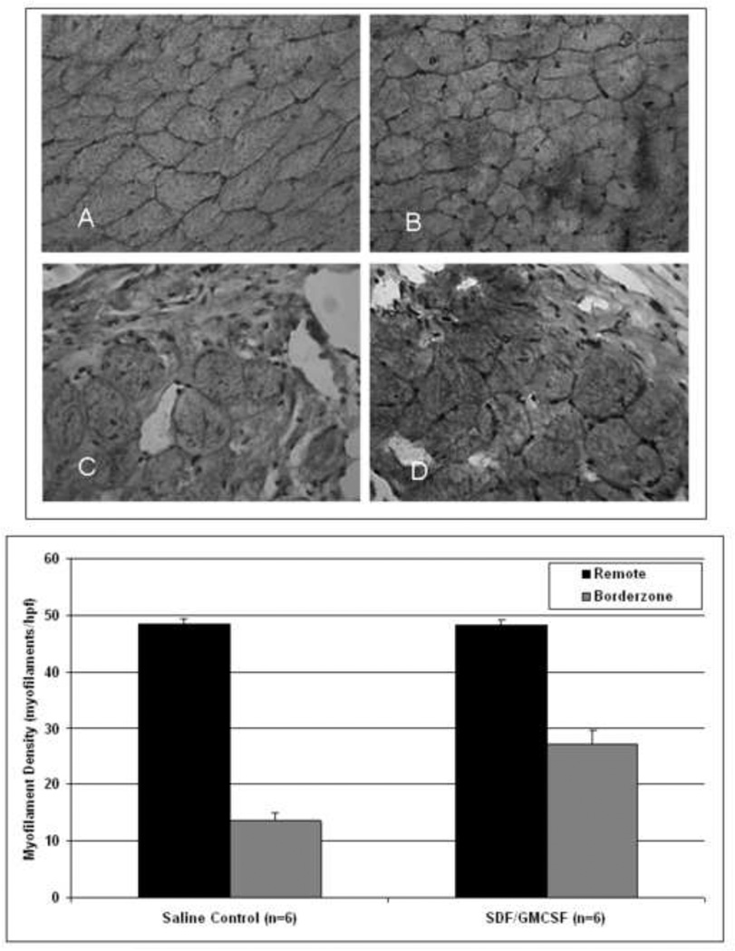
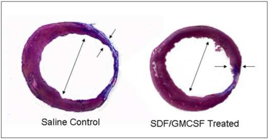
Findings: SDF/GMCSF therapy upregulated EPC density as shown by flow cytometry (0.12±0.02% vs. 0.06±0.01% circulating lymphocytes, p=0.005), 48hours following infarction. A marked increase in perfusion was evident eight weeks after therapy, utilising confocal angiography (5.02±1.7×10(-2)μm(3)blood/μm(3)myocardial tissue vs. 2.03±0.710(-2)μm(3)blood/μm(3)myocardial tissue, p=0.00004). Planimetric analysis demonstrated preservation of wall thickness (0.98±0.09mm vs. 0.67±0.06mm, p=0.003) and ventricular diameter (7.81±0.99mm vs. 9.41±1.1mm, p=0.03). Improved haemodynamic function was evidenced by echocardiography and PV analysis (ejection fraction: 56.4±18.1% vs. 25.3±15.6%, p=0.001; pre-load adjusted maximal power: 6.6±2.6mW/μl(2) vs. 2.7±1.4mW/μl(2), p=0.01).
Conclusion: Neovasculogenic therapy with GMCSF-mediated EPC upregulation and SDF-mediated EPC chemokinesis maybe an effective therapy for infarct modulation and preservation of myocardial function following acute myocardial infarction.
Copyright © 2010 Australasian Society of Cardiac and Thoracic Surgeons and the Cardiac Society of Australia and New Zealand. Published by Elsevier B.V. All rights reserved.
Figures
Similar articles
-
Neovasculogenic therapy to augment perfusion and preserve viability in ischemic cardiomyopathy.Ann Thorac Surg. 2006 May;81(5):1728-36. doi: 10.1016/j.athoracsur.2005.12.015. Ann Thorac Surg. 2006. PMID: 16631663
-
Transplantation of endothelial progenitor cells improves neovascularization and left ventricular function after myocardial infarction in a rat model.Basic Res Cardiol. 2008 Jan;103(1):69-77. doi: 10.1007/s00395-007-0685-9. Epub 2007 Nov 12. Basic Res Cardiol. 2008. PMID: 17999028
-
Stromal cell-derived factor and granulocyte-monocyte colony-stimulating factor form a combined neovasculogenic therapy for ischemic cardiomyopathy.J Thorac Cardiovasc Surg. 2005 Aug;130(2):321-9. doi: 10.1016/j.jtcvs.2004.11.041. J Thorac Cardiovasc Surg. 2005. PMID: 16077394
-
Granulocyte-colony stimulating factor therapy to induce neovascularization in ischemic heart disease.Dan Med J. 2012 Mar;59(3):B4411. Dan Med J. 2012. PMID: 22381094 Review.
-
SDF-1α as a therapeutic stem cell homing factor in myocardial infarction.Pharmacol Ther. 2011 Jan;129(1):97-108. doi: 10.1016/j.pharmthera.2010.09.011. Epub 2010 Oct 20. Pharmacol Ther. 2011. PMID: 20965212 Review.
Cited by
-
Oxygen-dependent quenching of phosphorescence used to characterize improved myocardial oxygenation resulting from vasculogenic cytokine therapy.J Appl Physiol (1985). 2011 May;110(5):1460-5. doi: 10.1152/japplphysiol.01138.2010. Epub 2011 Feb 3. J Appl Physiol (1985). 2011. PMID: 21292844 Free PMC article.
-
Myocardial tissue elastic properties determined by atomic force microscopy after stromal cell-derived factor 1α angiogenic therapy for acute myocardial infarction in a murine model.J Thorac Cardiovasc Surg. 2012 Apr;143(4):962-6. doi: 10.1016/j.jtcvs.2011.12.028. Epub 2012 Jan 20. J Thorac Cardiovasc Surg. 2012. PMID: 22264415 Free PMC article.
-
The State of Art of Regenerative Therapy in Cardiovascular Ischemic Disease: Biology, Signaling Pathways, and Epigenetics of Endothelial Progenitor Cells.Cells. 2020 Aug 11;9(8):1886. doi: 10.3390/cells9081886. Cells. 2020. PMID: 32796767 Free PMC article. Review.
-
Normalization of postinfarct biomechanics using a novel tissue-engineered angiogenic construct.Circulation. 2013 Sep 10;128(11 Suppl 1):S95-104. doi: 10.1161/CIRCULATIONAHA.112.000368. Circulation. 2013. PMID: 24030426 Free PMC article.
-
Sustained release of engineered stromal cell-derived factor 1-α from injectable hydrogels effectively recruits endothelial progenitor cells and preserves ventricular function after myocardial infarction.Circulation. 2013 Sep 10;128(11 Suppl 1):S79-86. doi: 10.1161/CIRCULATIONAHA.112.000343. Circulation. 2013. PMID: 24030424 Free PMC article.
References
-
- Boodhwani M, Sodha NR, Laham RJ, Sellke FW. The future of therapeutic myocardial angiogenesis. Shock. 2006 Oct;26(4):332–341. - PubMed
-
- Assmus B, Schachinger V, Teupe C, Britten M, Lehmann R, Dobert N, Grunwald F, Aicher A, Urbich C, Martin H, Hoelzer D, Dimmeler S, Zeiher AM. Transplantation of Progenitor Cells and Regeneration Enhancement in Acute Myocardial Infarction (TOPCARE-AMI) Circulation. 2002 Dec 10;106(24):3009–3017. - PubMed
-
- Woo YJ, Grand TJ, Berry MF, Atluri P, Moise MA, Hsu VM, Cohen J, Fisher O, Burdick J, Taylor M, Zentko S, Liao G, Smith M, Kolakowski S, Jayasankar V, Gardner TJ, Sweeney HL. Stromal cell-derived factor and granulocyte-monocyte colony-stimulating factor form a combined neovasculogenic therapy for ischemic cardiomyopathy. The Journal of thoracic and cardiovascular surgery. 2005 Aug;130(2):321–329. - PubMed
-
- Numaguchi Y, Sone T, Okumura K, Ishii M, Morita Y, Kubota R, Yokouchi K, Imai H, Harada M, Osanai H, Kondo T, Murohara T. The impact of the capability of circulating progenitor cell to differentiate on myocardial salvage in patients with primary acute myocardial infarction. Circulation. 2006 Jul 4;114(1 Suppl):I114–I119. - PubMed
-
- Simons M. Angiogenesis: where do we stand now? Circulation. 2005 Mar 29;111(12):1556–1566. - PubMed
Publication types
MeSH terms
Substances
Grants and funding
LinkOut - more resources
Full Text Sources
Medical
