

Human oocyte maturation is dependent on LH-stimulated accumulation of the epidermal growth factor-like growth factor, amphiregulin
- PMID: 20719813
- PMCID: PMC2939758
- DOI: 10.1093/humrep/deq212
Human oocyte maturation is dependent on LH-stimulated accumulation of the epidermal growth factor-like growth factor, amphiregulin
Abstract
Background: The LH surge promotes ovulation via activation of multiple signaling networks in the ovarian follicle. Studies in animal models have shown the importance of LH-induced activation of the epidermal growth factor (EGF)signaling network in critical peri-ovulatory events. We investigated the biological significance of regulatory mechanisms mediated by EGF-like growth factors during LH stimulation in humans.
Methods: We characterized the EGF signaling network in mature human ovarian follicles using in vivo and in vitro approaches. Amphiregulin (AREG) levels were measured in 119 follicular fluid (FF) samples from IVF/ICSI patients. Biological activity of human FF was assessed using in vitro oocyte maturation, cumulus expansion and cell mitogenic assays.
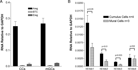
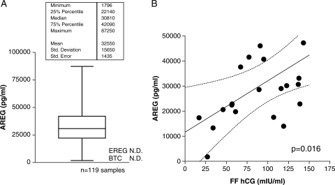
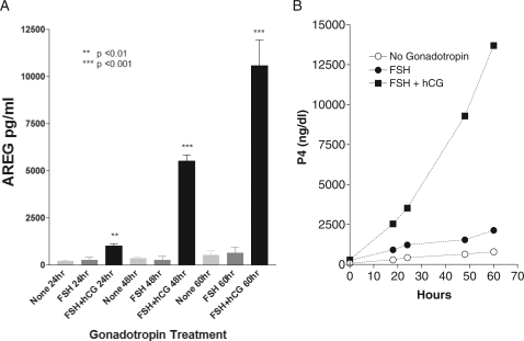
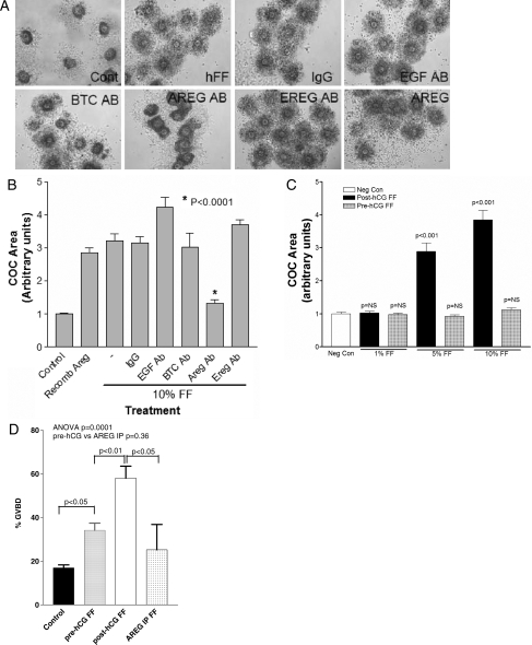
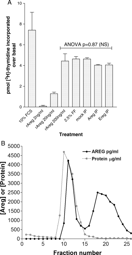
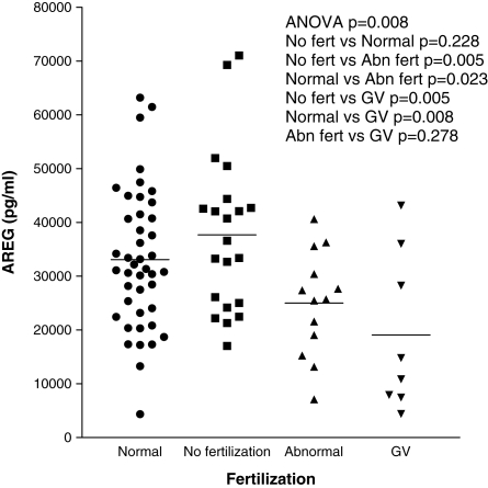
Results: AREG is the most abundant EGF-like growth factor accumulating in the FF of mature follicles of hCG-stimulated patients. No AREG was detected before the LH surge or before hCG stimulation of granulosa cells in vitro, demonstrating that the accumulation of AREG requires gonadotrophin stimulation. Epiregulin and betacellulin mRNA were detected in both human mural and cumulus granulosa cells, although at significantly lower levels than AREG. FF from stimulated follicles causes cumulus expansion and oocyte maturation in a reconstitution assay. Immunodepletion of AREG abolishes the ability of FF to stimulate expansion (P < 0.0001) and oocyte maturation (P < 0.05), confirming the biological activity of AREG. Conversely, mitogenic activity of FF remained after depletion of AREG, indicating that other mitogens accumulate in FF. FF from follicles yielding an immature germinal vesicle oocyte or from an oocyte that develops into an aberrant embryo contains lower AREG levels than that from follicles yielding a healthy oocyte (P = 0.008).
Conclusions: EGF-like growth factors play a role in critical peri-ovulatory events in humans, and AREG accumulation is a useful marker of gonadotrophin stimulation and oocyte competence.
Figures
Similar articles
-
Paracrine and autocrine regulation of epidermal growth factor-like factors in cumulus oocyte complexes and granulosa cells: key roles for prostaglandin synthase 2 and progesterone receptor.Mol Endocrinol. 2006 Jun;20(6):1352-65. doi: 10.1210/me.2005-0504. Epub 2006 Mar 16. Mol Endocrinol. 2006. PMID: 16543407
-
Amphiregulin promotes the maturation of oocytes isolated from the small antral follicles of the rhesus macaque.Hum Reprod. 2012 Aug;27(8):2430-7. doi: 10.1093/humrep/des158. Epub 2012 May 16. Hum Reprod. 2012. PMID: 22593432 Free PMC article.
-
Hormone-induced expression of tumor necrosis factor alpha-converting enzyme/A disintegrin and metalloprotease-17 impacts porcine cumulus cell oocyte complex expansion and meiotic maturation via ligand activation of the epidermal growth factor receptor.Endocrinology. 2007 Dec;148(12):6164-75. doi: 10.1210/en.2007-0195. Epub 2007 Sep 27. Endocrinology. 2007. PMID: 17901238
-
The epidermal growth factor network: role in oocyte growth, maturation and developmental competence.Hum Reprod Update. 2018 Jan 1;24(1):1-14. doi: 10.1093/humupd/dmx029. Hum Reprod Update. 2018. PMID: 29029246 Review.
-
Epidermal growth factor-like growth factors in the follicular fluid: role in oocyte development and maturation.Semin Reprod Med. 2009 Jan;27(1):52-61. doi: 10.1055/s-0028-1108010. Epub 2009 Feb 5. Semin Reprod Med. 2009. PMID: 19197805 Free PMC article. Review.
Cited by
-
New insights into the ovulatory process in the human ovary.Hum Reprod Update. 2025 Jan 1;31(1):21-47. doi: 10.1093/humupd/dmae027. Hum Reprod Update. 2025. PMID: 39331957 Free PMC article. Review.
-
Proteomic analysis of human follicular fluid from fertile women.Clin Proteomics. 2015 Mar 3;12(1):5. doi: 10.1186/s12014-015-9077-6. eCollection 2015. Clin Proteomics. 2015. PMID: 25838815 Free PMC article.
-
Activation of P2X7 Receptor Mediates the Abnormal Ovulation Induced by Chronic Restraint Stress and Chronic Cold Stress.Biology (Basel). 2024 Aug 15;13(8):620. doi: 10.3390/biology13080620. Biology (Basel). 2024. PMID: 39194558 Free PMC article.
-
Melatonin Alleviates the Suppressive Effect of Hypoxanthine on Oocyte Nuclear Maturation and Restores Meiosis via the Melatonin Receptor 1 (MT1)-Mediated Pathway.Front Cell Dev Biol. 2021 Apr 15;9:648148. doi: 10.3389/fcell.2021.648148. eCollection 2021. Front Cell Dev Biol. 2021. PMID: 33937242 Free PMC article.
-
Improved efficiency of microsurgical enucleated tripronuclear zygotes development and embryonic stem cell derivation by supplementing epidermal growth factor, brain-derived neurotrophic factor, and insulin-like growth factor-1.Stem Cells Dev. 2014 Mar 15;23(6):563-75. doi: 10.1089/scd.2013.0420. Epub 2014 Jan 24. Stem Cells Dev. 2014. PMID: 24261581 Free PMC article.
References
-
- Aaltonen J, Laitinen MP, Vuojolainen K, Jaatinen R, Horelli-Kuitunen N, Seppa L, Louhio H, Tuuri T, Sjoberg J, Butzow R, et al. Human growth differentiation factor 9 (GDF-9) and its novel homolog GDF-9B are expressed in oocytes during early folliculogenesis. J Clin Endocrinol Metab. 1999;84:2744–2750. doi:10.1210/jc.84.8.2744. - DOI - PubMed
-
- Adam RM, Borer JG, Williams J, Eastham JA, Loughlin KR, Freeman MR. Amphiregulin is coordinately expressed with heparin-binding epidermal growth factor-like growth factor in the interstitial smooth muscle of the human prostate. Endocrinology. 1999;140:5866–5875. doi:10.1210/en.140.12.5866. - DOI - PubMed
-
- Ashkenazi H, Cao X, Motola S, Popliker M, Conti M, Tsafriri A. Epidermal growth factor family members: endogenous mediators of the ovulatory response. Endocrinology. 2005;146:77–84. doi:10.1210/en.2004-0588. - DOI - PubMed
-
- Ben-Ami I, Freimann S, Armon L, Dantes A, Ron-El R, Amsterdam A. Novel function of ovarian growth factors: combined studies by DNA microarray, biochemical and physiological approaches. Mol Hum Reprod. 2006;12:413–419. doi:10.1093/molehr/gal045. - DOI - PubMed
-
- Buccione R, Vanderhyden BC, Caron PJ, Eppig JJ. FSH-induced expansion of the mouse cumulus oophorus in vitro is dependent upon a specific factor(s) secreted by the oocyte. Dev Biol. 1990;138:16–25. doi:10.1016/0012-1606(90)90172-F. - DOI - PubMed
Publication types
MeSH terms
Substances
Grants and funding
LinkOut - more resources
Full Text Sources
Other Literature Sources
