. 2010 Sep;8(9):1234-47.
doi: 10.1158/1541-7786.MCR-10-0233. Epub 2010 Aug 18.

Chk1-dependent constitutive phosphorylation of BLM helicase at serine 646 decreases after DNA damage

Affiliations

Chk1-dependent constitutive phosphorylation of BLM helicase at serine 646 decreases after DNA damage

Sarabpreet Kaur et al. Mol Cancer Res. 2010 Sep.

Abstract

BLM helicase, the protein mutated in Bloom syndrome, is involved in signal transduction cascades after DNA damage. BLM is phosphorylated on multiple residues by different kinases either after stress induction or during mitosis. Here, we have provided evidence that both Chk1 and Chk2 phosphorylated the NH(2)-terminal 660 amino acids of BLM. An internal region within the DExH motif of BLM negatively regulated the Chk1/Chk2-dependent NH(2)-terminal phosphorylation event. Using in silico analysis involving the Chk1 structure and its known substrate specificity, we predicted that Chk1 should preferentially phosphorylate BLM on serine 646 (Ser(646)). The prediction was validated in vitro by phosphopeptide analysis on BLM mutants and in vivo by usage of a newly generated phosphospecific polyclonal antibody. We showed that the phosphorylation at Ser(646) on BLM was constitutive and decreased rapidly after exposure to DNA damage. This resulted in the diminished interaction of BLM with nucleolin and PML isoforms, and consequently decreased BLM accumulation in the nucleolus and PML nuclear bodies. Instead, BLM relocalized to the sites of DNA damage and bound with the damage sensor protein, Nbs1. Mutant analysis confirmed that the binding to nucleolin and PML isoforms required Ser(646) phosphorylation. These results indicated that Chk1-mediated phosphorylation on BLM at Ser(646) might be a determinant for regulating subnuclear localization and could act as a marker for the activation status of BLM in response to DNA damage.


Figures

Figure 1
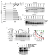
Figure 1. Chk1 and Chk2 phosphorylate BLM in the first 660 amino acids
A. Chk2 phosphorylated wildtype BLM. Recombinant Chk1 (WT or KD) was incubated with BLM (1–1417) (400 ng on left and 100 ng, 400 ng on right) in the presence of γ-32P ATP. The proteins were resolved by SDS-PAGE and detected by autoradiography. B. (Left) Schematic diagram of full-length BLM and its fragments (1–212), (191–660), (621–1041) and (1001–1417). (Right) Levels of BLM (1–212), BLM (191–660), BLM (621–1041) and BLM (1001–1417) as detected by Coomassie. Approximately 1 µg of protein was leaded in each lane. C. Interaction between in vitro translated S35-radiolabelled Chk1 and equal amounts (5 µg) of the glutathione-sepharose bound BLM fragments or GST alone. The amount of bound radioactivity was detected by autoradiography. D. Same as (C), except S35-radiolabelled Chk2 was used for interaction with BLM (1–1417). E. Chk1 phosphorylated BLM (1–212) and (1–660). Chk1-dependent kinase assays were carried out with 400 ng of BLM (1–212), BLM (191–660), BLM (621–1041) and BLM (1001–417). Arrows indicated the phosphorylated products. F. Same as (E), except Chk2 was used as the kinase to phosphorylate BLM fragments.
Figure 2
Figure 2. Amino acids within DExH motif negatively regulated the Chk1-mediated N-terminal phosphorylation of BLM
A. Schematic diagram and relative expression levels of wild type BLM and its derivatives. (Left) Full length BLM (1–1417) and its various fragments (1–660), (1–800), (1–900), (1–1006), (1–1041), (1–1211), (1–1292) and (661–800). The helicase, RQC and HRDC domains are indicated. (Right, top) The expression of the BLM (1–660), (1–800), (1–900), (1–1006), (1–41041), (1–1211), (1–31292) and (1–1417) fragments were determined by Coomassie. Approximately 1 µg of protein was leaded in each lane. B. In vitro phosphorylation of full-length BLM and C-terminal deletion fragments [i.e. BLM (1–660), (1–800), (1–900), (1–1006), (1–1041), (1–1211) and (1–1292)] (400 ng each) in presence of γ32P-ATP and recombinant Chk1. C. BLM (661–800) negatively regulated the phosphorylation of BLM (1–660). (Left) BLM (661–800) was expressed, purified and checked by Coomassie along with BLM (1–660) and GST. Approximately 1 µg of protein was leaded in each lane. (Middle) Chk1-dependent phosphorylation of BLM (1–660) (400 ng) was carried out either alone or in presence of increasing amounts (50 ng, 100 ng, 200 ng, 400 ng) of BLM (661–800). As control, phosphorylation was done for GST (400 ng), BLM (661–800) (400 ng) and BLM (1–660) (400 ng) in presence of GST (400 ng). (Right) Graph indicates the extent of inhibition of the phosphorylation of BLM (1–660) by GST, Chk1 or Chk2. The values are represented by mean with the standard deviation. D. BLM (661–800) inhibited BLM (1–600) phosphorylation even after preincubation with AMP-PNP. Chk1-dependent phosphorylation of BLM (1–660) (400 ng), alone or in presence of BLM (661–800) (400 ng) was carried out either without or after preincubation of BLM (661–800) with AMP-PNP (5 µM).
Figure 3
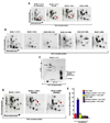
Figure 3. Chk1 phosphorylated BLM at Ser646 in vitro
A. Phosphopeptide maps of human BLM (1–1417) (produced either in S. cerevisiae or in E. coli) and BLM fragments (1–1041) and (1–660) phosphorylated in vitro by Chk1. Red arrow indicates a phosphopeptide present in all except BLM (1–660). The black arrows indicate the directions in which the phosphopeptides were separated by electrophoresis and chromatography in the first and second dimensions, respectively. B. Same as (A) except the following BLM derivatives were used: BLM (1–212), BLM (109–212), BLM (191–660), BLM (531–660), BLM (1–660) and BLM (1–1417). C. Phosphoaminoacid analysis of BLM (1417). The arrows indicate the directions during the chromatographic runs. The broken circles indicate the positions of co-migrating cold phosphoaminoacid standards. The position of the origin and products of the partial hydrolysis have been indicated. D. Phosphopeptide analysis of BLM (1–1041) and the two mutants BLM (1–1041) S646A and BLM (1–660) S646A. Arrow indicates the position of the phosphopeptide decreased in the mutants. E. Peptides (containing either wild type or mutant Ser646 residue) were phosphorylated with either the wild type or kinase dead recombinant Chk1. Bound radioactivity was quantitated by scintillation counting.
Figure 4
Figure 4. BLM phosphorylation at Ser646 decreased after DNAdamage
A. (Left) Cell cycle profile of A-15 and BS, both grown asynchronously, and A-15 treated with UCN-01 for 2 h. (Middle) Nuclear extracts (50 µg) from A-15, grown in the above two conditions, and BS (grown asynchronously) were subjected to SDS-PAGE and western blotting to determine the level of BLM and hsp90. (Right) Nuclear extracts (1 mg) from A-15 and BS were immunoprecipitated with BLM antibody and the immoprecipitates subjected to western blotting with either BLM or pSer646BLM antibody. B. BLM was immunoprecipitated from nuclear extracts (1 mg) obtained from BS and A-15 cells (−HU, +HU, PW) with anti-BLM antibody. The immunoprecipitates were probed with either BLM (top) or pSer646BLM antibody (bottom). Immunoprecipitates from A-15 cells were additionally incubated with Calf Intestinal Alkaline Phosphatase (CIAP, 10 units) for 30 min before western blotting to check for the specificity of pSer646BLM signal under-HU condition. (*) indicated a cross-reactive band. C. Immunoprecipitaion with anti-BLM antibody was carried out on nuclear extracts isolated from A-15 cells obtained from different conditions. The immunoprecipitates were probed for the presence of BLM, pSer646BLM, nucleolin, PML isoforms and Nbs1. D. Immunoprecipitaion with anti-pSer646BLM antibody was carried out on nuclear extracts isolated from A-15 cells obtained from different conditions. The immunoprecipitates were probed for the presence of pSer646BLM, BLM and nucleolin.
Figure 5
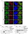
Figure 5. Ser646 phosphorylation of BLM regulates its localization in PML NBs and nucleolin
A. A-15 cells were either left untreated (−HU) or treated with HU (+HU) for 12 h. Immunofluorescence was carried out with antibodies against (a, d) BLM/pSer646BLM, (b) pSer646BLM/PML, (c) pSer646BLM/nucleolin (e) BLM/Nbs1. Nucleus is stained by DAPI. Combined indicates the merged image from the red and green channel. The numbers indicate the percentage of cells having similar colocalization. B–C. Immunoprecipitaions were carried out with either anti-nucleolin (B) or anti-PML (C) antibody on cell extracts (1 mg) obtained after transfecting Cos cells with either pcDNA3 Flag BLM (wildtype) or pcDNA3 Flag BLM (S646A). The immunoprecipitates were probed for the presence of nucleolin and Flag i.e BLM (for B) or PML isoforms and Flag i.e. BLM (for C).

Similar articles

Cited by

References

    1. Cimprich KA, Cortez D. ATR: an essential regulator of genome integrity. Nat Rev Mol Cell Biol. 2008;9:616–627. - PMC - PubMed
    1. Hurley PJ, Bunz F. ATM and ATR: components of an integrated circuit. Cell Cycle. 2007;6:414–417. - PubMed
    1. Bartek J, Lukas J. Chk1 and Chk2 kinases in checkpoint control and cancer. Cancer Cell. 2003;3:421–429. - PubMed
    1. Hickson ID. RecQ helicases: caretakers of the genome. Nat Rev Cancer. 2003;3:169–178. - PubMed
    1. Sharma S, Doherty KM, Brosh RM., Jr Mechanisms of RecQ helicases in pathways of DNA metabolism and maintenance of genomic stability. Biochem J. 2006;398:319–337. - PMC - PubMed

Publication types