. 2010 Nov;84(21):11210-8.
doi: 10.1128/JVI.01211-10. Epub 2010 Aug 18.

Swine influenza H1N1 virus induces acute inflammatory immune responses in pig lungs: a potential animal model for human H1N1 influenza virus

Affiliations

Swine influenza H1N1 virus induces acute inflammatory immune responses in pig lungs: a potential animal model for human H1N1 influenza virus

Mahesh Khatri et al. J Virol. 2010 Nov.

Abstract

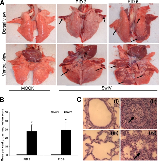
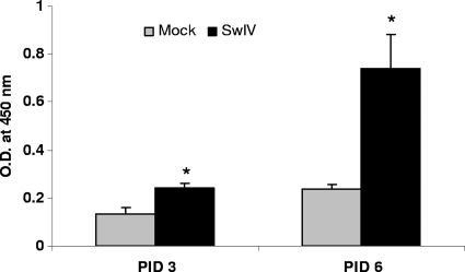
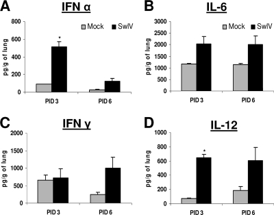
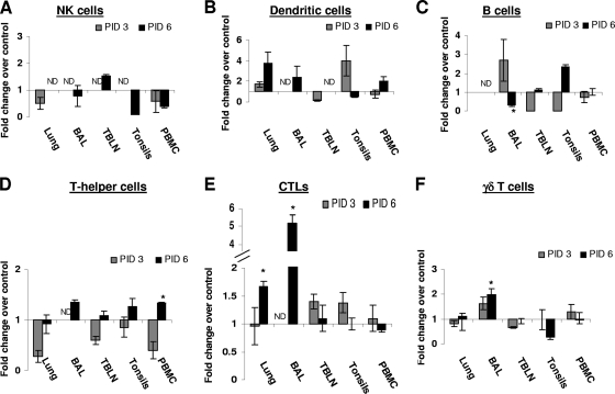
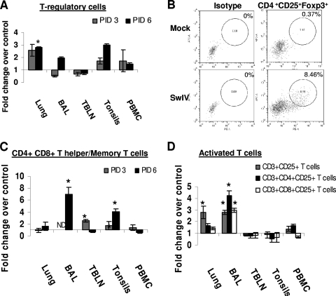
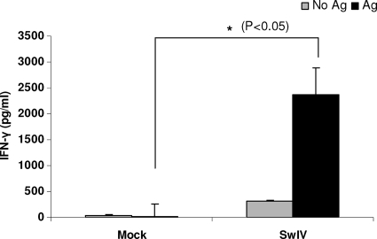
Pigs are capable of generating reassortant influenza viruses of pandemic potential, as both the avian and mammalian influenza viruses can infect pig epithelial cells in the respiratory tract. The source of the current influenza pandemic is H1N1 influenza A virus, possibly of swine origin. This study was conducted to understand better the pathogenesis of H1N1 influenza virus and associated host mucosal immune responses during acute infection in humans. Therefore, we chose a H1N1 swine influenza virus, Sw/OH/24366/07 (SwIV), which has a history of transmission to humans. Clinically, inoculated pigs had nasal discharge and fever and shed virus through nasal secretions. Like pandemic H1N1, SwIV also replicated extensively in both the upper and lower respiratory tracts, and lung lesions were typical of H1N1 infection. We detected innate, proinflammatory, Th1, Th2, and Th3 cytokines, as well as SwIV-specific IgA antibody in lungs of the virus-inoculated pigs. Production of IFN-γ by lymphocytes of the tracheobronchial lymph nodes was also detected. Higher frequencies of cytotoxic T lymphocytes, γδ T cells, dendritic cells, activated T cells, and CD4+ and CD8+ T cells were detected in SwIV-infected pig lungs. Concomitantly, higher frequencies of the immunosuppressive T regulatory cells were also detected in the virus-infected pig lungs. The findings of this study have relevance to pathogenesis of the pandemic H1N1 influenza virus in humans; thus, pigs may serve as a useful animal model to design and test effective mucosal vaccines and therapeutics against influenza virus.


Figures

FIG. 1.
FIG. 1.
Gross lung lesions in pigs infected with SwIV OH07. (A) Five-week-old pigs were mock infected or infected with SwIV. At PIDs 3 and 6, extensive areas of purple-red consolidation indicative of severe pneumonia were observed in lungs of virus-infected pigs. (B) Mean gross lung lesions from all the six lung lobes were scored on the basis of percent pneumonic lesions. Virus-infected pigs had significantly higher (P < 0.05) mean lung lesions than mock-infected pigs at both PIDs 3 and 6. Each bar indicates the mean lung lesion scores of three pigs ± standard error of the mean (SEM). Asterisk indicates significant differences (P < 0.05) in virus-infected pig lungs compared to mock-infected control pigs. Arrows indicate the purple-red consolidation in virus-infected pig lungs. A representative control and SwIV-infected pig lung is shown. (C) Microscopic lung lesions in pigs infected with SwIV OH07. Control uninfected pig lung showing normal bronchial epithelial lining and absence of inflammatory cell infiltrates and debris in the lumen (panel i). SwIV-induced bronchiolitis at PID 3 characterized by bronchial epithelial necrosis, numerous intraepithelial leukocytes, and subepithelial and peribronchial mononuclear inflammatory cell infiltrations. The bronchial lumen contains desquamated bronchial epithelium and dead and dying inflammatory cells, mainly neutrophils and proteinic debris, indicated by an arrow (panel ii). Panel iii shows control uninfected lung, showing normal alveolar walls, clear air space, and absence of exudation into the alveolar space. Panel iv shows SwIV-induced exudative interstitial pneumonia at PID 3, illustrating the thickened alveolar walls containing intravascular and extravascular inflammatory cells, indicated by an arrow. The alveolar spaces are collapsed and contain proteinic debris and an increased population of inflammatory cells. Hematoxylin and eosin stain. Magnification, ×200. Representative control and SwIV-infected pig lung sections are shown. Similar histological changes were detected in pig lungs of another independent experiment.
FIG. 2.
FIG. 2.
SwIV-specific IgA antibody response in the lungs of experimentally infected pigs. On PIDs 3 and 6, virus-specific IgA antibody production in lungs of mock- and SwIV-infected pigs was analyzed by ELISA. Each bar indicates mean OD450 ± SEM for three pigs. Asterisk indicates significant differences (P < 0.05) in virus-infected pig lungs compared to mock-infected control pigs. A similar trend in the virus-specific IgA antibody response was also found in another independent experiment.
FIG. 3.
FIG. 3.
Cytokine production in pig lungs infected with SwIV OH07. Five-week-old pigs were mock infected or infected intranasally and intratracheally with SwIV. At PIDs 3 and 6, cytokine production in lung lysate of mock- and virus-infected pigs was analyzed: innate (IFN-α) (A), proinflammatory (IL-6) (B), Th1 IFN-γ (C), and IL-12 cytokines (D) by ELISA. Each bar indicates mean concentrations of cytokines ± SEM for three pigs. Asterisk indicates significant differences (P < 0.05) in virus-infected pig lungs compared to mock-infected control pigs. A similar trend in the cytokine response was detected in another independent experiment.
FIG. 4.
FIG. 4.
Comparison of innate immune cells (NK cells and DC), B cells, and T lymphocyte subsets in different tissues of mock-infected and SwIV-infected pigs. Five-week-old pigs were mock-infected or infected with SwIV. At PIDs 3 and 6, MNC isolated from the lungs, BAL fluid, TBLN, tonsils, and PBMC were immunostained with appropriate antibodies or its isotype control MAb and then analyzed by flow cytometry for NK cells (A), DC cells (B), B cells (C), T-helper cells (D), CTLs (E), and γδ T cells (F). Data shown are the fold increase or decrease in the mean (n = 3 pigs/group) percentage of indicated pig immune cells of SwIV versus those of mock-infected pigs. A similar trend in the frequency of immune cells was detected in another independent experiment.
FIG. 5.
FIG. 5.
Frequency of T regulatory cells, T-helper/memory cells, and activated T cells in SwIV-infected pigs. Five-week-old pigs were mock infected or infected with SwIV. At PIDs 3 and 6, MNC isolated from the lungs, BAL fluid, TBLN, tonsils, and PBMC were immunostained with appropriate antibodies or its isotype control MAb and then analyzed by flow cytometry for T regulatory cells (CD4+ CD25+ Foxp3+) (A and B), CD4+ CD8+ T helper/memory T cells (C), and activated T cells (D) at PID 6. Data shown are the fold increase or decrease in the mean (n = 3 pigs/group) percentage of immune cells of SwIV over those of mock-infected pigs. A representative dot plot of results for a mock- and an SwIV-infected pig showing the frequency of Tregs in the pig lungs at PID 6 is shown (B). Asterisk indicates significant differences (P < 0.05) in the SwIV- versus mock-infected pig immune cells. A similar trend in these immune cells was detected in another independent experiment.
FIG. 6.
FIG. 6.
IFN-γ production by in vivo SwIV OH07 primed pig lymphocytes of TBLN upon ex vivo restimulation. TBLN-MNC of mock- and SwIV-infected pigs at PID 6 were restimulated ex vivo in the presence of inactivated SwIV antigens. IFN-γ production in the supernatant was measured by ELISA. Each bar indicates the mean IFN-γ concentration ± SEM for three pigs. Asterisk indicates significant differences (P < 0.05) in the amount of IFN-γ secreted by SwIV- versus mock-infected pig cells. Similar results were also found in another independent experiment.

Similar articles

Cited by

References

    1. Azevedo, M. S., L. Yuan, S. Pouly, A. M. Gonzales, K. I. Jeong, T. V. Nguyen, and L. J. Saif. 2006. Cytokine responses in gnotobiotic pigs after infection with virulent or attenuated human rotavirus. J. Virol. 80:372-382. - PMC - PubMed
    1. Barbe, F., K. Atanasova, and K. Van Reeth. 2010. Cytokines and acute phase proteins associated with acute swine influenza infection in pigs. Vet. J. doi:10.1016/j.tvjl.2009.12.012. - DOI - PMC - PubMed
    1. Reference deleted.
    1. Basta, S., C. P. Carrasco, S. M. Knoetig, R. C. Rigden, H. Gerber, A. Summerfield, and K. C. McCullough. 2000. Porcine alveolar macrophages: poor accessory or effective suppressor cells for T-lymphocytes. Vet. Immunol. Immunopathol. 77:177-190. - PubMed
    1. Brookes, S. M., A. Nunez, B. Choudhury, M. Matrosovich, S. C. Essen, D. Clifford, M. J. Slomka, G. Kuntz-Simon, F. Garcon, B. Nash, A. Hanna, P. M. Heegaard, S. Queguiner, C. Chiapponi, M. Bublot, J. M. Garcia, R. Gardner, E. Foni, W. Loeffen, L. Larsen, K. Van Reeth, J. Banks, R. M. Irvine, and I. H. Brown. 2010. Replication, pathogenesis and transmission of pandemic (H1N1) 2009 virus in non-immune pigs. PLoS One 5:e9068. - PMC - PubMed

MeSH terms