. 2010 Nov;84(21):11189-99.
doi: 10.1128/JVI.01075-10. Epub 2010 Aug 18.

The F gene of the Osaka-2 strain of measles virus derived from a case of subacute sclerosing panencephalitis is a major determinant of neurovirulence

Affiliations

The F gene of the Osaka-2 strain of measles virus derived from a case of subacute sclerosing panencephalitis is a major determinant of neurovirulence

Minoru Ayata et al. J Virol. 2010 Nov.

Abstract

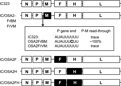
Measles virus (MV) is the causative agent for acute measles and subacute sclerosing panencephalitis (SSPE). Although numerous mutations have been found in the MV genome of SSPE strains, the mutations responsible for the neurovirulence have not been determined. We previously reported that the SSPE Osaka-2 strain but not the wild-type strains of MV induced acute encephalopathy when they were inoculated intracerebrally into 3-week-old hamsters. The recombinant MV system was adapted for the current study to identify the gene(s) responsible for neurovirulence in our hamster model. Recombinant viruses that contained envelope-associated genes from the Osaka-2 strain were generated on the IC323 wild-type MV background. The recombinant virus containing the M gene alone did not induce neurological disease, whereas the H gene partially contributed to neurovirulence. In sharp contrast, the recombinant virus containing the F gene alone induced lethal encephalopathy. This phenotype was related to the ability of the F protein to induce syncytium formation in Vero cells. Further study indicated that a single T461I substitution in the F protein was sufficient to transform the nonneuropathogenic wild-type MV into a lethal virus for hamsters.


Figures

FIG. 1.
FIG. 1.
Schematic diagram of the genome of the recombinant MVs. The protein-coding regions (N, P, M, F, H, and L) of the IC323 strain are shown as open boxes. The M, F, and H protein-coding regions derived from the Osaka-2 strain are shown as filled boxes. The sequence of the P gene end of the two sibling viruses (OSA2FrBM and OSA2FrVM) of the Osaka-2 strain and the extents of the transcriptional read-through at the P-M gene junction are indicated.
FIG. 2.
FIG. 2.
(A) Plaque titration of the CD-VLP of recombinant MV containing the M gene of either OSA2FrV (IC/OSA2FrVM; black bars) or OSA2FrB (IC/OSA2FrBM; gray bars). Three different kinds of cell lines were used for the titration. The means ± standard deviations for triplicate samples are shown. (B and C) Growth kinetics of the recombinant MVs containing the M gene. B95a cells were infected with each recombinant MV at a multiplicity of infection of 0.01 PFU/cell. At indicated time points, culture fluids were collected, an equal volume of medium was added, and cells were frozen and thawed once, clarified by centrifugation, and then titrated. Filled circles, IC323; open triangles, IC/OSA2FrBM; open circles, IC/OSA2FrVM. Data shown are for infectious cell-free viruses released into the culture medium (B) and cell-associated viruses prepared by the freezing and thawing of infected cells (C).
FIG. 3.
FIG. 3.
Infectious cell-free virus production by the recombinant MVs. B95a cells were infected with each recombinant MV at a multiplicity of infection of 0.01 PFU/cell. At 72 h p.i., the virus released into the culture fluid (cell-free) or the cell-associated virus prepared by the freezing and thawing of infected cells (cell-associated) was titrated on B95a cells. The virus titer in cells infected with the IC323 strain was set to 100%. Bars indicate the means with standard deviations for triplicate samples.
FIG. 4.
FIG. 4.
Histopathological findings of the hippocampus. Neuronal loss and degenerated neurons were observed with different extents of severity in fields CA1 through CA3 of hamsters inoculated with the IC/OSA2FH, IC/OSA2F, IC/OSA2H, IC/F:OSA2F-ext, IC/F:T461I, and IC/F:Y442D (B to E, G, and H), whereas no lesion was detected in the brain of the hamsters inoculated with the wild-type IC323, IC/F:OSA2-cyt, IC/F:L197I, or IC/F:E478G virus (A, F, I, and J). In hamsters inoculated with the IC/OSA2H virus (D), neuronal damage was restricted to the CA1 field of the hippocampus. Proliferation and activation of astroglia and microglia were also noted in the affected areas. Inoculated viruses were the following: IC323 (hamster 262; showed no symptoms and was sacrificed at 70 days p.i.) (A); IC/OSA2FH (hamster 290; showed symptoms at 4 days p.i. and was sacrificed at 6 days p.i.) (B); IC/OSA2F (hamster 176; showed symptoms at 3 days p.i. and was sacrificed at 6 days p.i.) (C); IC/OSA2H (hamster 187; showed symptoms at 7 days p.i. and was sacrificed at 12 days p.i.) (D); IC/F:OSA2-ext (hamster 237; showed symptoms at 3 days p.i. and died at 5 days p.i.) (E); IC/F:OSA2-cyt (hamster 245; showed no symptoms and was sacrificed at 280 days p.i.) (F); IC/F:T461I (hamster 275; showed symptoms at 5 days p.i. and was sacrificed at 6 days p.i.) (G); IC/F:Y442D (hamster 268; showed symptoms at 4 days p.i. and was sacrificed at 168 days p.i.) (H); IC/F:L197I (hamster 258; showed no symptoms and was sacrificed at 70 days p.i.) (I); IC/F:E478G (hamster 278; showed no symptoms and was sacrificed at 70 days p.i.) (J). CA1, CA2, CA3, and DG (dentate gyrus) fields are indicated in panel A. Scale bar, 500 μm. Panels A, B, and G to J show the hippocampus area of the left hemisphere, and panels C to F show the hippocampus area of the right hemisphere of the brain. The arrows indicate the abnormal appearance of fields CA1 through CA3 due to the necrosis of pyramidal cells.
FIG. 5.
FIG. 5.
Schematic diagram of the MV F protein and constructs. HRA and HRB, two highly conserved heptad repeat domains, are shown; regions of signal peptide and fusion peptide are indicated as shaded boxes. TM, transmembrane domain (black box). Arrows indicate amino acids different from the those of the IC323 strain. For the F and H coexpression experiments by transfection, the fragments were inserted into a eukaryotic expression vector, pME18S. For the recombinant MV constructions, plasmids were prepared by replacing the whole F gene fragment from the basic plasmid containing the IC323 viral genome.
FIG. 6.
FIG. 6.
Syncytium formation induced by a different kind of mutant F protein. Vero cells were cotransfected with plasmid DNA encoding the H and F proteins of MV. The H gene was derived from the IC323 strain while the F gene was from one of the mutant F genes. At 24 h p.i., cells were fixed and stained with a monoclonal antibody to the H protein and fluorescein isothiocyanate-conjugated anti-mouse antibody. (A) IC/F:L197I. (B) IC/F:Y442D. (C) IC/F:T461I. (D) IC/F:E478G. Arrows in panel C indicate syncytia formed in cells cotransfected with the F gene containing the T461I substitution. No syncytia were found in cells cotransfected with other F genes.

Similar articles

Cited by

References

    1. Albrecht, P., and H. P. Schumacher. 1971. Neurotropic properties of measles virus in hamsters and mice. J. Infect. Dis. 124:86-93. - PubMed
    1. Albrecht, P., T. Burnstein, M. J. Klutch, H. T. Hicks, and F. A. Ennis. 1977. Subacute sclerosing panencephalitis: experimental infection in primates. Science 195:64-66. - PubMed
    1. Ayata, M., A. Hirano, and T. C. Wong. 1989. Structural defect linked to nonrandom mutations in the matrix gene of Biken strain subacute sclerosing panencephalitis virus defined by cDNA cloning and expression of chimeric genes. J. Virol. 63:1162-1173. - PMC - PubMed
    1. Ayata, M., K. Hayashi, T. Seto, R. Murata, and H. Ogura. 1998. The matrix gene expression of subacute sclerosing panencephalitis (SSPE) virus (Osaka-1 strain): a comparison of two sibling viruses isolated from different lobes of an SSPE brain. Microbiol. Immunol. 42:773-780. - PubMed
    1. Ayata, M., K. Komase, M. Shingai, I. Matsunaga, Y. Katayama, and T. C. Wong. 2002. Mutations affecting transcriptional termination in the P gene end of subacute sclerosing panencephalitis viruses. J. Virol. 76:13062-13068. - PMC - PubMed

Publication types

Substances

LinkOut - more resources