. 2010 Nov;84(21):11113-23.
doi: 10.1128/JVI.01183-10. Epub 2010 Aug 18.

Epstein-Barr virus nuclear antigen 1 Hijacks the host kinase CK2 to disrupt PML nuclear bodies

Affiliations

Epstein-Barr virus nuclear antigen 1 Hijacks the host kinase CK2 to disrupt PML nuclear bodies

Nirojini Sivachandran et al. J Virol. 2010 Nov.

Abstract

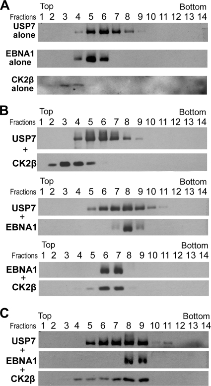
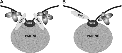
Latent Epstein-Barr virus (EBV) infection is an important causative factor in the development of several cancers, including nasopharyngeal carcinoma (NPC). The one EBV protein expressed in the nucleus of NPC cells, EBNA1, has been shown to disrupt promyelocitic leukemia (PML) nuclear bodies (NBs) by inducing the degradation of PML proteins, leading to impaired DNA repair and increased cell survival. Although EBNA1-mediated PML disruption is likely to be an important factor in the development of NPC, little is known about its mechanism. We now show that an interaction between EBNA1 and the host CK2 kinase is crucial for EBNA1 to disrupt PML bodies and degrade PML proteins. EBNA1 increases the association of CK2 with PML proteins, thereby increasing the phosphorylation of PML proteins by CK2, a modification that is known to trigger the polyubiquitylation and degradation of PML. The interaction between EBNA1 and CK2 is direct and occurs through the β regulatory subunit of CK2 and EBNA1 amino acids 387 to 394. The binding of EBNA1 to the host ubiquitin specific protease USP7 has also been shown to be important for EBNA1-mediated PML disruption. We show that EBNA1 also increases the occupancy of USP7 at PML NBs and that CK2 and USP7 bind independently and simultaneously to EBNA1 to form a ternary complex. The combined results indicate that EBNA1 usurps two independent cellular pathways to trigger the loss of PML NBs.


Figures

FIG. 1.
FIG. 1.
EBNA1-mediated PML degradation requires CK2 and involves increased PML phosphorylation. (A) CNE2 cells and CNE2E cells were transfected with siRNA against CK2α (siCK2α) or GFP (siGFP; negative control) and then stained for CK2α and PML. Images were captured using the same exposure time for both cell lines. (B) Quantification of the number of PML NBs per cell in each of the samples in A. P values are <0.01 for siGFP and siCK2 in CNE2 and <0.001 for siGFP and siCK2 in CNE2E. (C) Equal amounts of cell lysates from panel A were analyzed by Western blotting with the indicated antibodies, where “−” samples are treated with siGFP and “+” samples are treated with siCK2α. (D) CNE2 and CNE2E were treated with 10 μM emodin (in dimethyl sulfoxide [DMSO]; +) or DMSO alone (−) or for 3 days, and then equal amount of cell lysates were analyzed by Western blotting with the indicated antibodies, including one that recognizes only PML phosphorylated at S517 (p517). (E) Equal amount of CNE2 and CNE2E cell lysates were analyzed by Western blotting with the indicated antibodies. (F) CNE2 and CNE2E cells were stained with antibodies against total PML and PML phosphorylated at serine 517 (p517). Images were captured using the same exposure time for both cell lines.
FIG. 2.
FIG. 2.
EBNA1 directly interacts with CK2β. (A) Purified EBNA1 was incubated with purified GST-tagged CK2α or CK2β or with GST alone at equimolar ratios and then mixed with glutathione-Sepharose. After washing, proteins were eluted with glutathione and analyzed by SDS-PAGE and colloidal Coomassie blue staining. The samples shown are the input protein mixture (I; 3% of the total), the flowthrough that was not retained on the resin (F; 3% of the total), and protein that was retained by the resin and eluted (E; 50% of the total). The positions of EBNA1, CK2α-GST, CK2β-GST, and molecular weight markers (M) in kilodaltons are indicated. (B) Purified EBNA1, CK2α, or CK2β proteins were analyzed by glycerol gradient sedimentation individually (top panels) or after preincubation of equal molar ratios of EBNA1 and CK2β or CK2α (middle or bottom panels). Equal volume fractions were collected from top of each gradient, and equal volumes of each fractions were analyzed by Western blotting with antibodies against EBNA1, CK2α, or CK2β.
FIG. 3.
FIG. 3.
Mapping of CK2-binding region of EBNA1 by coimmunoprecipitation. (A) Schematic representation of EBNA1 and the EBNA1 mutants used in the present study, showing the glycine and arginine-rich regions (G/R), the glycine and alanine-rich region (G/A), the nuclear localization signal (NLS), and the DNA-binding domain (DNA BD). Note that the EBNA1 used in the present study contains a short version of the G/A repeat. The ability of the EBNA1 proteins to bind endogenous CK2 by coimmunoprecipitation is also indicated (as determined in panels B and C). (B and C) 293T cells were transfected with plasmids expressing the indicated FLAG-tagged EBNA1 protein or FLAG-tagged LacZ, and tagged proteins were immunoprecipitated from cell lysates using anti-FLAG resin. Proteins recovered from the FLAG resin were analyzed by Western blotting with antibodies against EBNA1 (B) or the FLAG epitope (C), CK2α (B and C), CK2β (C), and USP7 (C). Western blots are also shown for 10% of the starting cell lysates (Input).
FIG. 4.
FIG. 4.
The EBNA1 CK2-binding mutant fails to disrupt PML NBs. (A) CNE2 cells were transiently transfected with a plasmid expressing EBNA1 or EBNA1 mutant Δ387-394, then stained for EBNA1 and PML 24 h posttransfection. Both EBNA1-expressing (red) cells and nonexpressing cells are shown within the same image. (B) The numbers of PML NBs per cell were counted 24 h after expression of EBNA1 or Δ387-394 and compared to control cells transfected with the empty expression plasmid (OriP). (C) CNE2 cells were transiently transfected with a plasmid expressing EBNA1, Δ387-394, Δ395-450, or no protein (OriP) and, 48 h later, equal amounts of cell lysates were analyzed by Western blotting with the indicated antibodies.
FIG. 5.
FIG. 5.
EBNA1 increases association of CK2β and USP7 with PML NBs. (A) EBNA1 mutants Δ387-394 or Δ395-450 were expressed in CNE2 by transient transfection and, 12 h later, the cells were stained for EBNA1 and PML. (B) CNE2 cells were transiently transfected with the EBNA1 expression plasmid and, 12 h later, the cells were stained for EBNA1, USP7, and PML with specific antibodies directly conjugated to Alexa Fluor dyes. Overlays of USP7 with PML and EBNA1 with PML are also shown in which USP7 and EBNA1 have been colorized red and PML is green. (C) Quantification of the percentage of PML NBs that have associated USP7 foci with or without EBNA1 expression (P < 0.001). (D to F) CNE2 cells were transfected with a plasmid expressing EBNA1 (+) or the empty plasmid (−) and, 14 h later, immunoprecipitations were performed from nuclear lysates using anti-USP7 (D) or anti-PML antibody (E and F). IgG is a negative control in which immunoprecipitations were performed with rabbit IgG beads. Samples were Western blotted with the indicated antibodies. One-fifteenth of the starting nuclear extracts (input) is also shown. In panel E, the blot was initially probed for USP7 (bottom panel) and then stripped and reprobed for PML (top panel). Due to incomplete stripping, some residual USP7 is still visible, as indicated in the top panel.
FIG. 6.
FIG. 6.
EBNA1, USP7, and CK2β form a ternary complex. Glycerol gradient sedimentation was performed with purified EBNA1, CK2β, and USP7 on 10 to 20% glycerol gradients. Equal volume fractions were collected from top of each gradient and equal volumes of each fraction were analyzed by Western blotting with antibodies against each protein (indicate on the left of each panel). Fraction 14 is the pellet fraction from the bottom of the tube. (A) The sedimentation of USP7, EBNA1 or CK2β proteins was analyzed individually (top, middle, and bottom panels, respectively). (B) Equal molar ratios of USP7 and CK2β (top panel), USP7 and EBNA1 (middle panel), or EBNA1 and CK2β (bottom panel) were preincubated then analyzed by glycerol gradient sedimentation. (C) Equal molar ratios of USP7, CK2β, and EBNA1 were preincubated and then analyzed by glycerol gradient sedimentation.
FIG. 7.
FIG. 7.
Models of the CK2-EBNA1-USP7 ternary complex. An EBNA1 dimer (black) associated with a PML NB is shown where the black oval represents the dimerized dimerization/DNA-binding domain. CK2 (α and β) and USP7 proteins are shown bound to the extended EBNA1 sequences outside of the dimerization domain. (A) CK2 and USP7 associate with the same monomer within the EBNA1 dimer. (B) CK2 and USP7 interact with opposite monomers within the EBNA1 dimer due to steric hindrance.

Similar articles

Cited by

References

    1. Adamson, A. L., and S. Kenney. 2001. Epstein-Barr virus immediate-early protein BZLF1 is SUMO-1 modified and disrupts promyelocytic leukemia bodies. J. Virol. 75:2388-2399. - PMC - PubMed
    1. Ahn, J. H., and G. S. Hayward. 1997. The major immediate-early proteins IE1 and IE2 of human cytomegalovirus colocalize with and disrupt PML-associated nuclear bodies at very early times in infected permissive cells. J. Virol. 71:4599-4613. - PMC - PubMed
    1. Ambinder, R. F., M. Mullen, Y. Chang, G. S. Hayward, and S. D. Hayward. 1991. Functional domains of Epstein-Barr nuclear antigen EBNA-1. J. Virol. 65:1466-1478. - PMC - PubMed
    1. Appel, K., P. Wagner, B. Boldyreff, O. G. Issinger, and M. Montenarh. 1995. Mapping of the interaction sites of the growth suppressor protein p53 with the regulatory beta-subunit of protein kinase CK2. Oncogene 11:1971-1978. - PubMed
    1. Bell, P., P. M. Lieberman, and G. G. Maul. 2000. Lytic but not latent replication of Epstein-Barr virus is associated with PML and induces sequential release of nuclear domain 10 proteins. J. Virol. 74:11800-11810. - PMC - PubMed

Publication types

MeSH terms