. 2010 Nov;84(21):11418-28.
doi: 10.1128/JVI.01131-10. Epub 2010 Aug 18.

A conserved domain in the coronavirus membrane protein tail is important for virus assembly

Affiliations

A conserved domain in the coronavirus membrane protein tail is important for virus assembly

Ariel L Arndt et al. J Virol. 2010 Nov.

Abstract

Coronavirus membrane (M) proteins play key roles in virus assembly, through M-M, M-spike (S), and M-nucleocapsid (N) protein interactions. The M carboxy-terminal endodomain contains a conserved domain (CD) following the third transmembrane (TM) domain. The importance of the CD (SWWSFNPETNNL) in mouse hepatitis virus was investigated with a panel of mutant proteins, using genetic analysis and transient-expression assays. A charge reversal for negatively charged E(121) was not tolerated. Lysine (K) and arginine (R) substitutions were replaced in recovered viruses by neutrally charged glutamine (Q) and leucine (L), respectively, after only one passage. E121Q and E121L M proteins were capable of forming virus-like particles (VLPs) when coexpressed with E, whereas E121R and E121K proteins were not. Alanine substitutions for the first four or the last four residues resulted in viruses with significantly crippled phenotypes and proteins that failed to assemble VLPs or to be rescued into the envelope. All recovered viruses with alanine substitutions in place of SWWS residues had second-site, partially compensating, changes in the first TM of M. Alanine substitution for proline had little impact on the virus. N protein coexpression with some M mutants increased VLP production. The results overall suggest that the CD is important for formation of the viral envelope by helping mediate fundamental M-M interactions and that the presence of the N protein may help stabilize M complexes during virus assembly.


Figures
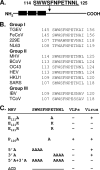
FIG. 1.
FIG. 1.
M protein conserved domain and mutants. (A) A linear schematic of the M protein illustrating the relative positions of the three TM domains (black boxes) and the position of the CD in the tail. (B) Alignment of CDs from representative coronaviruses. Full-length amino acid sequences from transmissible gastroenteritis virus (TGEV), feline coronavirus (FeCoV), human coronavirus 229E, human coronavirus NL63, mouse hepatitis virus (MHV), bovine coronavirus (BCoV), human coronavirus OC43, porcine hemagglutinating encephalomyelitis virus (HEV), human coronavirus HKU1, SARS-CoV, infectious bronchitis virus (IBV), and turkey coronavirus (TCoV) were aligned by using CLUSTAL W (25). (C) Mutations introduced into the MHV CD, with + and − symbols used to indicate VLP production and virus recovery for each mutant.
FIG. 2.
FIG. 2.
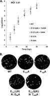
Growth properties of single-substitution mutant viruses. (A) Mouse 17Cl1 cells were infected with mutant viruses at an MOI of 0.01 PFU/cell. Titers were determined by plaque assay on L2 cells at the indicated times. Data points are shown for all viruses, but the growth curve is included for only the WT virus. Error bars and exponential growth curves are as described in the Materials and Methods. The estimated doubling time was ∼0.62 h, and the saturation parameter was 1.5% per h. (B) Plaque characteristics were determined for the indicated viruses in L2 cells. Changes in the recovered viruses with positive-charge (R and K) substitutions and corresponding noncompensating changes in the N gene are indicated.
FIG. 3.
FIG. 3.
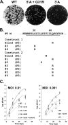
Growth properties of 5′A and 3′A mutant viruses. (A) Plaque characteristics of WT, 5′A+G31R, and 3′A viruses were analyzed in mouse L2 cells. (B) Summary of second-site changes in the TM1 of recovered viruses from two independent virus constructions, designated 1 and 2, are shown under the sequence of WT TM1 (underlined). Assigned numbers for recovered plaque-purified isolates and the passage numbers when analyzed are indicated. (C) Growth kinetics experiments were performed with mouse 17Cl1 cells infected at the indicated MOIs. Titers were determined by plaque assay on L2 cells at the indicated times. Data represent averages from two independent growth kinetic experiments as described for Fig. 2 and in Materials and Methods. Estimated doubling times were ∼0.62 h for WT virus, ∼0.79 h for all 5′A viruses except L35P (∼1.2 h), and 1 h for the 3′A virus.
FIG. 4.
FIG. 4.
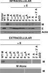
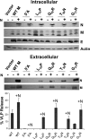
Effects of CD mutations on VLP production. 293T cells were transfected with pCAGGS vectors containing WT or mutant M genes singly or in combination with pCAGGS containing the WT E gene. Control empty vector (vector) was analyzed in parallel. Intracellular cell lysates and pelleted extracellular VLPs were analyzed by SDS-PAGE and Western blotting using antibodies against M, E, or actin as an internal loading control. The entire VLP pellet and 6% of the total intracellular fractions were analyzed.
FIG. 5.
FIG. 5.
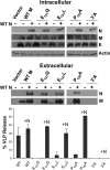
N protein enhancement of VLP production. 293T cells were transfected as indicated with pCAGGS vectors containing WT or mutant M genes in combination with pCAGGS-E and pCAGGS-N where indicated. Intracellular and extracellular VLP fractions were analyzed by SDS-PAGE and Western blotting as indicated in Fig. 4. The entire extracellular pellet and 6% of the total intracellular fraction were analyzed. Protein bands were quantified by densitometric scanning and analyzed using ImageQuant software. VLP release was calculated as the percentage of the extracellular M compared to total M (intracellular plus extracellular) protein. The data represent averages and standard deviations from two experiments.
FIG. 6.
FIG. 6.
VLP analysis of 5′A M mutants with second-site changes in TM1 coexpressed with the N protein. 293T cells were transfected with WT and mutant genes in pCAGGS vectors. M genes were expressed with E in the absence and presence of the N gene. Intracellular and extracellular fractions were analyzed by SDS-PAGE and Western blotting. Protein bands from the entire VLP pellet and 6% of total cytoplasmic lysates were quantified, and release was calculated as described for Fig. 5. Error bars represent the standard deviations from two independent experiments.
FIG. 7.
FIG. 7.
Rescue of CD mutants into VLPs. 293T cells were transfected with the pCAGGS vector containing WT or mutant M proteins as indicated. Cells were labeled for 20 h at 4 h after transfection with Expre35S35S labeling mixture. Intracellular lysates (Intra) and extracellular media (Extra) were divided in half and immunoprecipitated with monoclonal J1.3 or polyclonal F88 antibodies, which recognize only WT and both WT and A2A3 proteins, respectively. Samples were analyzed by SDS-PAGE and autoradiography.
FIG. 8.
FIG. 8.
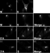
S protein colocalization with WT and mutant M proteins. 293T cells were transfected with pCAGGS vectors containing WT or mutant M proteins and the S gene. Cells were fixed at 12 h after transfection and analyzed by immunofluorescence using mouse and goat antibodies against the M and S proteins, respectively. Alexa Fluor 488-conjugated mouse and Alexa Fluor 594-conjugated goat secondary antibodies were used to visualize the localizations of the M and S proteins, respectively. Singly expressed M and S proteins are shown in the top two panels. Colocalizations of the M and S proteins are shown in the merged images on the right.

Similar articles

Cited by

References

    1. Barcena, M., G. T. Oostergetel, W. Bartelink, F. G. Faas, A. Verkleij, P. J. Rottier, A. J. Koster, and B. J. Bosch. 2009. Cryo-electron tomography of mouse hepatitis virus: insights into the structure of the coronavirion. Proc. Natl. Acad. Sci. U. S. A. 106:582-587. - PMC - PubMed
    1. Bos, E. C., W. Luytjes, H. V. van der Meulen, H. K. Koerten, and W. J. Spaan. 1996. The production of recombinant infectious DI-particles of a murine coronavirus in the absence of helper virus. Virology 218:52-60. - PMC - PubMed
    1. Chen, C. Y., C. K. Chang, Y. W. Chang, S. C. Sue, H. I. Bai, L. Riang, C. D. Hsiao, and T. H. Huang. 2007. Structure of the SARS coronavirus nucleocapsid protein RNA-binding dimerization domain suggests a mechanism for helical packaging of viral RNA. J. Mol. Biol. 368:1075-1086. - PMC - PubMed
    1. Cologna, R., J. F. Spagnolo, and B. G. Hogue. 2000. Identification of nucleocapsid binding sites within coronavirus-defective genomes. Virology 277:235-249. - PMC - PubMed
    1. Corse, E., and C. E. Machamer. 2000. Infectious bronchitis virus E protein is targeted to the Golgi complex and directs release of virus-like particles. J. Virol. 74:4319-4326. - PMC - PubMed

Publication types

LinkOut - more resources