. 2010 Nov;84(21):11245-54.
doi: 10.1128/JVI.00372-10. Epub 2010 Aug 18.

Poxvirus complement control proteins are expressed on the cell surface through an intermolecular disulfide bridge with the viral A56 protein

Affiliations

Poxvirus complement control proteins are expressed on the cell surface through an intermolecular disulfide bridge with the viral A56 protein

Brian C DeHaven et al. J Virol. 2010 Nov.

Abstract

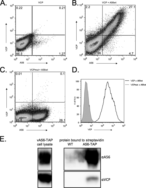
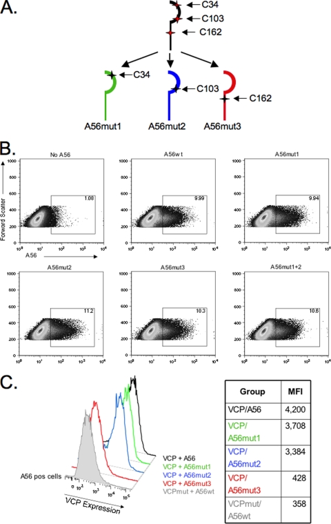
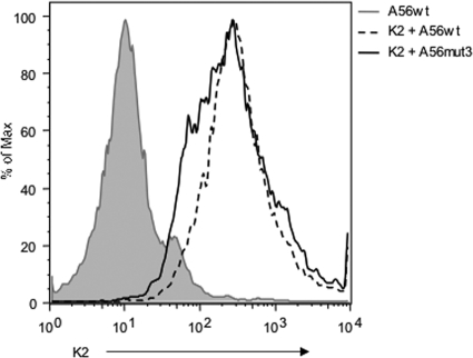
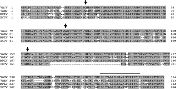
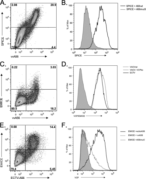
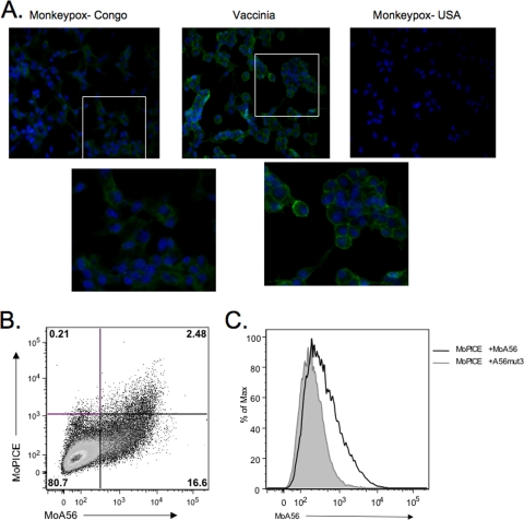
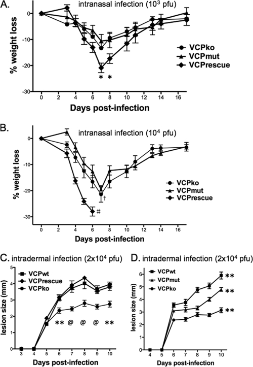
The vaccinia virus (VACV) complement control protein (VCP) is an immunomodulatory protein that is both secreted from and expressed on the surface of infected cells. Surface expression of VCP occurs though an interaction with the viral transmembrane protein A56 and is dependent on a free N-terminal cysteine of VCP. Although A56 and VCP have been shown to interact in infected cells, the mechanism remains unclear. To investigate if A56 is sufficient for surface expression, we transiently expressed VCP and A56 in eukaryotic cell lines and found that they interact on the cell surface in the absence of other viral proteins. Since A56 contains three extracellular cysteines, we hypothesized that one of the cysteines may be unpaired and could therefore form a disulfide bridge with VCP. To test this, we generated a series of A56 mutants in which each cysteine was mutated to a serine, and we found that mutation of cysteine 162 abrogated VCP cell surface expression. We also tested the ability of other poxvirus complement control proteins to bind to VACV A56. While the smallpox homolog of VCP is able to bind VACV A56, the ectromelia virus (ECTV) VCP homolog is only able to bind the ECTV homolog of A56, indicating that these proteins may have coevolved. Surface expression of poxvirus complement control proteins may have important implications in viral pathogenesis, as a virus that does not express cell surface VCP is attenuated in vivo. This suggests that surface expression of VCP may contribute to poxvirus pathogenesis.


Figures

FIG. 1.
FIG. 1.
VCP and A56 interact on transfected and infected cells. VCP, VCPmut, and A56wt were transfected into 293T cells either alone or in combination. After 48 h, the cells were collected and stained under nonpermeabilized conditions for FACS analysis with an anti-VCP polyclonal antibody and anti-56 MAb LC10. (A) VCP is not expressed on the cell surface when transfected alone. (B and C) VCP, but not VCPmut, is expressed on the cell surface in the presence of A56. (D) Levels of VCP expression on A56-positive cells. The black solid line represents cells transfected with VCP and A56wt; the gray shaded area represents cells transfected with VCPmut and A56wt. (E) VCP is present after pulling down A56-TAP with streptavidin from cells infected with virus expressing TAP-tagged A56 (A56-TAP). Western blots were probed with anti-A56 and anti-VCP antibodies to show the presence of the indicated proteins. Boxed areas on the left represent blots of the input cell lysate from cells infected with virus expressing A56-TAP. Boxed areas on the right represent blots of the proteins pulled down with streptavidin from cells infected with virus expressing untagged A56 (WT) or from cells infected with virus expressing TAP-tagged A56.
FIG. 2.
FIG. 2.
A56 mutagenesis and VCP surface expression. (A) Schematic of the cysteine pattern in the ectodomain of A56 and the three cysteine mutant A56 proteins. (B) Expression levels of A56wt versus A56mut1, A56mut2, A56mut3, and A56mut1+2. (C) Cells positive for A56 expression were gated and analyzed for VCP expression. Mean fluorescence intensity (MFI) levels are listed in the table to the right of the figure. VCP was cotransfected with the indicated A56 construct and stained with anti-VCP MAb 3F11, anti-A56 rabbit Ab, and appropriate secondary Abs and analyzed.
FIG. 3.
FIG. 3.
A56mut3 is able to bind K2. 293T cells were transfected with A56wt alone or with K2 plus A56wt and A56mut3. The cells were then stained with anti-K2 MAb 4A11-4A3 and the rabbit anti-A56Ab. A56-positive cells were gated and used to create the histogram.
FIG. 4.
FIG. 4.
Comparison of orthopoxvirus A56 genes. The protein sequences of vaccinia virus (VACV-WR) A56 and the homologs in variola virus (VARV-Bangladesh), monkeypox virus (MPXV-Zaire), and ectromelia virus (ECTV-Moscow) were downloaded from poxvirus.org and aligned using BLAST. Shaded residues indicate identical amino acids to vaccinia virus, and dashes represent missing amino acids. Conserved cysteines in the ectodomain are indicated in bold and with an arrow. The putative transmembrane domain is marked with the black line above the residues.
FIG. 5.
FIG. 5.
SPICE and EMICE are also expressed on the cell surface in the presence of A56. SPICE is expressed on cells cotransfected with vaccinia virus A56wt. (A) Scatter plot of SPICE cotransfected with A56wt. (B) Histogram of SPICE expression on A56-positive cells. The black line is SPICE transfected with vaccinia virus A56wt, and the shaded gray area is SPICE transfected with A56mut3. (C) Scatter plot of EMICE transfected with VACV A56. (D) Histogram of CCP expression on virus-infected cells. RK-13 cells were infected with ECTV, VACV, or vv-VCPko and then stained with a polyclonal anti-VCP antibody. Shown is a histogram of surface staining due to EMICE (black line), VACV (dotted line), or vv-VCPko (gray shaded area). (E) Scatter plot of EMICE cotransfected with ECTV A56. (F) Histogram of EMICE expression of cells cotransfected with ECTV A56 (black line), VACV A56 (dotted line), or VACV A56mut3 (gray shaded area).
FIG. 6.
FIG. 6.
MoPICE is expressed on the cell surface at low levels. (A) Immunofluorescence of infected cells. BSC-40 cells were infected for 18 h with MPXV-Congo, VACV, or a CCP-minus strain of MPXV, MPXV-USA, and fixed and stained using anti-VCP MAb 2E5 and fluorescent secondary Ab. To better visualize the cell distribution of VCP or MoPICE, the white box indicates the area of the image that was enlarged (shown in the second row of images), but unaltered. (B) Scatter plot of MoPICE cotransfected with MPXV A56. (C) Histogram of MoPICE expression on A56-positive cells. MoPICE was cotransfected with MPXV-A56 (black line) or VACV A56mut3 (gray shaded area), and cells were then stained for FACS with polyclonal rabbit anti-VCP and anti-A56 MAb.
FIG. 7.
FIG. 7.
Wild-type VCP is needed for full virulence in mice. Our panel of recombinant viruses was studied in 6- to 8-week-old female C57BL/6 mice either after intranasal (A and B) or intradermal (C and D) inoculation. (A and B) Average weight loss of 6-week-old mice (n = 5 for all groups) infected with vaccinia virus encoding VCPrescue, VCPmut, or VCPko at 103 PFU (A) or 104 PFU (B). #, indicates that all mice infected with vv-VCPrescue died; †, a single mouse in the group infected with vv-VCPko died; *, significant difference (P < 0.05) on the days indicated between VCPrescue and VCPko and between VCPrescue and VCPmut (unpaired Student's t test). (C and D) Lesion diameters in 6- to 8-week-old mice inoculated with 2 × 10⁴ PFU of VCPrescue, VCPwt, or VCPko (C) or VCPwt, VCPko, or VCPmut (D). For panel C, data points represent the mean lesion diameter ± standard error of the mean of 10 infected ears per group (two ears per mouse). P values for the difference between VCPko and VCPwt and between VCPko and VCPrescue for each day are as follows: *, P < 0.05; **, P < 0.01; @, P < 0.001 (unpaired Student's t test). For panel D, data points represent the mean lesion diameter ± standard error of the mean of 20 (VCPko), 14 (VCPwt), or 10 (VCPmut) infected ears per group (two ears per mouse). Differences in lesion size between the groups of mice were statistically significant (P < 0.01; two-way ANOVA).

Similar articles

Cited by

References

    1. Benhnia, M. R., M. M. McCausland, J. Moyron, J. Laudenslager, S. Granger, S. Rickert, L. Koriazova, R. Kubo, S. Kato, and S. Crotty. 2009. Vaccinia virus extracellular enveloped virion neutralization in vitro and protection in vivo depend on complement. J. Virol. 83:1201-1215. - PMC - PubMed
    1. Bernet, J., J. Mullick, Y. Panse, P. B. Parab, and A. Sahu. 2004. Kinetic analysis of the interactions between vaccinia virus complement control protein and human complement proteins C3b and C4b. J. Virol. 78:9446-9457. - PMC - PubMed
    1. Blue, C. E., O. B. Spiller, and D. J. Blackbourn. 2004. The relevance of complement to virus biology. Virology 319:176-184. - PMC - PubMed
    1. Brown, C. K., P. C. Turner, and R. W. Moyer. 1991. Molecular characterization of the vaccinia virus hemagglutinin gene. J. Virol. 65:3598-3606. - PMC - PubMed
    1. Brum, L. M., P. C. Turner, H. Devick, M. T. Baquero, and R. W. Moyer. 2003. Plasma membrane localization and fusion inhibitory activity of the cowpox virus serpin SPI-3 require a functional signal sequence and the virus encoded hemagglutinin. Virology 306:289-302. - PubMed

Publication types

LinkOut - more resources