. 2010 Oct 1;21(19):3487-96.
doi: 10.1091/mbc.E10-02-0123. Epub 2010 Aug 18.

Sprouty proteins inhibit receptor-mediated activation of phosphatidylinositol-specific phospholipase C

Affiliations

Sprouty proteins inhibit receptor-mediated activation of phosphatidylinositol-specific phospholipase C

Simge Akbulut et al. Mol Biol Cell. .

Abstract

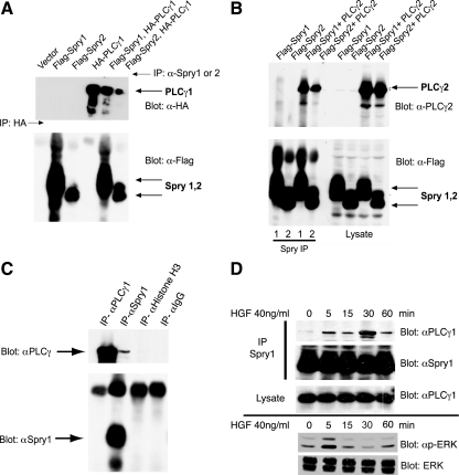
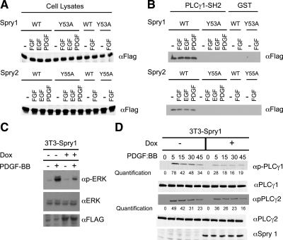
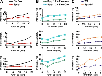
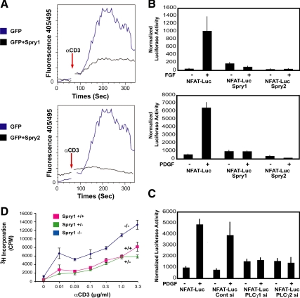
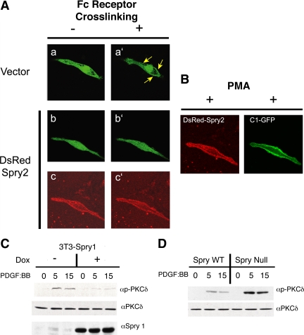
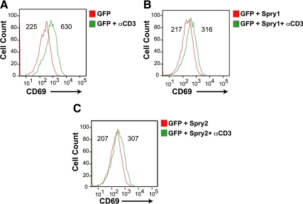
Sprouty (Spry) proteins are negative regulators of receptor tyrosine kinase signaling; however, their exact mechanism of action remains incompletely understood. We identified phosphatidylinositol-specific phospholipase C (PLC)-γ as a partner of the Spry1 and Spry2 proteins. Spry-PLCγ interaction was dependent on the Src homology 2 domain of PLCγ and a conserved N-terminal tyrosine residue in Spry1 and Spry2. Overexpression of Spry1 and Spry2 was associated with decreased PLCγ phosphorylation and decreased PLCγ activity as measured by production of inositol (1,4,5)-triphosphate (IP(3)) and diacylglycerol, whereas cells deficient for Spry1 or Spry1, -2, and -4 showed increased production of IP(3) at baseline and further increased in response to growth factor signals. Overexpression of Spry 1 or Spry2 or small-interfering RNA-mediated knockdown of PLCγ1 or PLCγ2 abrogated the activity of a calcium-dependent reporter gene, suggesting that Spry inhibited calcium-mediated signaling downstream of PLCγ. Furthermore, Spry overexpression in T-cells, which are highly dependent on PLCγ activity and calcium signaling, blocked T-cell receptor-mediated calcium release. Accordingly, cultured T-cells from Spry1 gene knockout mice showed increased proliferation in response to T-cell receptor stimulation. These data highlight an important action of Spry, which may allow these proteins to influence signaling through multiple receptors.


Figures

Figure 1.
Figure 1.
PLCγ1 and PLCγ2 coimmunoprecipitate with Spry1 and Spry2. (A) HEK293 cells were transfected with plasmids expressing epitope-tagged Spry and PLCγ as indicated. Spry1 and Spry2 were immunoprecipitated with anti-Spry1 or anti-Spry2 antibody. HA-PLCγ1 was immunoprecipitated (lane 4) with anti-HA antibody. The immunoprecipitates were probed with anti-HA antibody to detect coimmunoprecipitated PLCγ1 (top) and with anti-FLAG antibody to detect Spry1 and Spry2 (bottom). (B) HEK293 cells were cotransfected with cDNAs encoding FLAG-tagged Spry1 or FLAG-tagged Spry2 and PLCγ2 as indicated. After 48 h, lysates were prepared, and immunoprecipitations were performed using specific anti-Spry1 or anti-Spry2 antibody (left). Total cell lysate (50 μg) was resolved in 10% SDS-polyacrylamide gel electrophoresis gel and probed with anti-PLCγ2 and anti-FLAG antibodies to show the expression level of the transfectants (right). (C) mIMCD3 cells growing in media containing 10% FBS were lysed and subjected to immunoprecipitation with anti-Spry1 and anti-PLCγ1 antibodies or IgG or anti histone H3 controls, followed by immunoblotting with anti-Spry1 or PLCγ1. (D) Murine IMCD3 cells were serum starved for 18 h followed by HGF (40 ng/ml) stimulation for the indicated time. Endogenous Spry1 and PLCγ1 interaction was demonstrated by immunoprecipitation with anti-Spry1 antibody and immunoblotting for PLCγ1. Total cell lysate was blotted with PLCγ1 to show the endogenous level of PLCγ1 in the cell lysates after stimulation with HGF. Bottom, lysate from a second experiment was immunoblotted for activated phosphorylated (p)-ERK and total ERK to show the time course of activation of signaling in response to HGF.
Figure 2.
Figure 2.
Sprouty–PLCγ1 interaction requires an N-terminal conserved tyrosine residue of Sprouty. (A) 293T cells were transfected with either wild type or mutant Spry1 or Spry 2 as indicated. Thirty-six hours after transfection, cells were starved with 0.2% serum-containing medium for 12 h, and cells were stimulated with indicated growth factors for 10 min. Aliquots (50 μg) of whole cell lysate from unstimulated or stimulated cells were subjected to immunoblotting with anti-FLAG antibody to visualize Spry1 (top) and Spry2 (bottom) expression. (B) 293T cells were transfected with FLAG-tagged wild-type (WT) Spry1 or Spry2 or N-terminal tyrosine mutants as indicated. The cells were starved with 0.2% FBS-containing medium for 12 h and were stimulated with indicated growth factors for 10 min. The cell lysates from stimulated or unstimulated cells were incubated with 20 μg of GST or GST-PLCγ1-SH2-C fusion proteins. The GST-bound proteins were eluted in SDS loading buffer, and the bound Spry was detected by immunoblotting with anti-FLAG antibody. (C) Spry1 expression in a stable 3T3 cell line was induced by addition of doxycycline to starvation media containing 0.2% FBS and then stimulated with PDGF BB (20 ng/ml) for 15 min. Cell lysates were probed for total and phospho-ERK. (D) 3T3 cells were stimulated for increasing periods of time with PDGF BB (20 ng/ml) in the presence or absence of doxycycline (Dox) to induce Spry1 expression. Cell lysates were immunoblotted for total and phosphorylated PLCγ1 and PLCγ2 as indicated.
Figure 3.
Figure 3.
Spry inhibits inositol phosphate (IP) production after receptor stimulation. (A) NIH 3T3 cells cultured in the presence of [3H]inositol were serum starved and stimulated with PDGF BB (20 ng/ml) for increasing periods in the presence or absence of doxycycline (Dox) to induce Spry1 expression. Lipids were collected from the cell lysates, and radiolabeled IPs were fractionated and counted by liquid scintillation. Inositol phosphate levels are expressed as a percentage of total radioactive inositol incorporation. (B) Spry1,2,4 null or control adenovirus-GFP fibroblasts were serum starved and treated with PDGF BB (20 ng/ml) for the indicated times and assayed for IPs. (C) A bone marrow mast cell line derived from a wild-type or Spry2−/− mouse was treated with IgE followed by dinitrophenol-human serum albumin to cross-link Fc receptors. Cell lysates were prepared at the indicated times and assayed for IPs. All experiments were performed three times (see Supplemental Table 1, A–C) with similar results obtained.
Figure 4.
Figure 4.
Sprouty inhibits intracellular calcium mobilization and signaling in T-cells and fibroblasts. (A) Jurkat T-cells were cotransfected with a GFP expression vector (1 μg) along with either empty pcDNA (3 μg) or Spry1 or Spry2 expression vector (3 μg) as indicated. The cells were then loaded with indo-1 and after baseline measurements of 60 s and stimulated with anti-CD3 (3 mg/ml) antibody. Measurements were taken for up to 300 s. Intracellular calcium mobilization was analyzed by fluorescence-activated cell sorting analysis by monitoring the fluorescence emission ratio of the indo-1 bound to Ca2+ versus the free form at 405 and 495 nM, respectively. The experiment was performed three times with similar results obtained. (B) Spry1-inducible NIH 3T3 cells were transiently cotransfected with a reporter containing an NFAT-responsive element (5 μg) and Renilla (50 ng), starved for 24 h in media containing 0.2% FBS, and stimulated either with bFGF (20 ng/ml; top) or PDGF BB (20 ng/ml; bottom) for 4h. Spry1 was induced by doxycycline addition to the starvation media; Spry2 was expressed by transient transfection of a Spry2 expression plasmid in cells cultured in the absence of doxycycline. After growth factor addition, equal quantities of cell extracts (50 μg) were assayed using the Dual-Luciferase Assay kit (Promega), normalizing luciferase activity to Renilla luciferase activity. The experiments were repeated three times, and similar results were obtained. Luc, luciferase. (C) NIH 3T3 cells were treated with siGENOME SMARTpool directed against PLCγ1 or PLCγ2 for 24 h followed by transient transfection with NFAT luciferase reporter (5 μg) and Renilla (50 ng) plasmids by using FuGENE Transfection Reagent (GE Healthcare) in duplicate. Serum-starved (0.2% FBS-containing serum media) cells were either left unstimulated or stimulated with PDGF BB (20 ng/ml) for 4 h. Luciferase activity was assayed as described above. (D) T-cells from Spry1−/− mice were cultured in triplicate with the indicated amount of anti-CD3 antibody in combination with anti-CD28 (1 μg/ml) antibody for 72 h, and [3H]thymidine incorporation was quantified by liquid scintillation counting.
Figure 5.
Figure 5.
Spry2 prevents relocalization of a DAG reporter protein to the plasma membrane and inhibits signaling downstream of DAG. (A) Fluorescent images of RBL-2H3 mast cells transfected with a DAG binding reporter protein construct (PKCγ-C1-GFP) without (a and a′) or with (b and b′) transfection of Spry2 before (a and b) and after (a′ and b′) cross-linking of the IgE receptors with DNP-BSA. c and c′ show DsRed Spry2 expression in the mast cell presented in b and b′. (B) The mast cell from b and b′ was treated with PMA showing membrane localization of DsRed Spry2 and PKCγ-C1-GFP after stimulation. Sixteen vector-transfected cells and 13 Spry2-transfected cells were analyzed as described above with results presented in Table 1. (C) NIH 3T3 cells engineered to express Spry1 under doxycycline control treated overnight with doxycycline in starvation media, treated with PDGF BB (20 ng/ml) for the indicated times, and then cell lysates were immunoblotted with antibodies directed against PKCδ and phospho-threonine 505-modified PKCδ. (D) Sprouty1,2,4 null or control fibroblasts were serum starved and treated with PDGF BB (20 ng/ml) for the indicated times. Cell lysates were immunoblotted for PKCδ and phospho-threonine 505-PKCδ. WT, wild type.
Figure 6.
Figure 6.
Spry suppresses induction of CD69, an MAP-kinase dependent marker in T-cells. Jurkat cells were transfected with GFP alone (A), with GFP and Spry1 (B), or with GFP and Spry2 (C) and left unstimulated or stimulated for 16 h with anti-CD3; stained with phycoerythrin-conjugated anti-CD69 expression, and analyzed by flow cytometry. Histograms of cell number versus fluorescence intensity are plotted, and mean fluorescence intensity of each curve is presented. The experiments were repeated three times, and similar results were obtained.

Similar articles

Cited by

References

    1. Abe M., Naski M. C. Regulation of sprouty expression by PLCγ and calcium-dependent signals. Biochem. Biophys. Res. Commun. 2004;323:1040–1047. - PubMed
    1. Ayada T., Taniguchi K., Okamoto F., Kato R., Komune S., Takaesu G., Yoshimura A. Sprouty4 negatively regulates protein kinase C activation by inhibiting phosphatidylinositol 4,5-biphosphate hydrolysis. Oncogene. 2009;28:1076–1088. - PubMed
    1. Barker S. A., Caldwell K. K., Pfeiffer J. R., Wilson B. S. Wortmannin-sensitive phosphorylation, translocation, and activation of PLCgamma1, but not PLCgamma2, in antigen-stimulated RBL-2H3 mast cells. Mol. Biol. Cell. 1998;9:483–496. - PMC - PubMed
    1. Basson M. A., et al. Sprouty1 is a critical regulator of GDNF/RET-mediated kidney induction. Dev. Cell. 2005;8:229–239. - PubMed
    1. Basson M. A., Watson-Johnson J., Shakya R., Akbulut S., Hyink D., Costantini F. D., Wilson P. D., Mason I. J., Licht J. D. Branching morphogenesis of the ureteric epithelium during kidney development is coordinated by the opposing functions of GDNF and Sprouty1. Dev. Biol. 2006;299:466–477. - PubMed

Publication types

MeSH terms

Substances

LinkOut - more resources