. 2010 Nov;299(5):F1094-110.
doi: 10.1152/ajprenal.00056.2010. Epub 2010 Aug 18.

Fibroblast growth factor-10 signals development of von Brunn's nests in the exstrophic bladder

Affiliations

Fibroblast growth factor-10 signals development of von Brunn's nests in the exstrophic bladder

Rocky Eastman Jr et al. Am J Physiol Renal Physiol. 2010 Nov.

Abstract

von Brunn's nests have long been recognized as precursors of benign lesions of the urinary bladder mucosa. We report here that von Brunn's nests are especially prevalent in the exstrophic bladder, a birth defect that predisposes the patient to formation of bladder cancer. Cells of von Brunn's nest were found to coalesce into a stratified, polarized epithelium which surrounds itself with a capsule-like structure rich in types I, III, and IV collagen. Histocytochemical analysis and keratin profiling demonstrated that nested cells exhibited a phenotype similar, but not identical, to that of urothelial cells of transitional epithelium. Immunostaining and in situ hybridization analysis of exstrophic tissue demonstrated that the FGF-10 receptor is synthesized and retained by cells of von Brunn's nest. In contrast, FGF-10 is synthesized and secreted by mesenchymal fibroblasts via a paracrine pathway that targets basal epithelial cells of von Brunn's nests. Small clusters of 10pRp cells, positive for both FGF-10 and its receptor, were observed both proximal to and inside blood vessels in the lamina propria. The collective evidence points to a mechanism where von Brunn's nests develop under the control of the FGF-10 signal transduction system and suggests that 10pRp cells may be the original source of nested cells.


Figures

Fig. 1.
Fig. 1.
Structure of von Brunn's nests in exstrophic bladder. AC: Masson's trichrome stain. D: immunohistochemistry with antibodies specific for type IV collagen. A and C: bar = 50 μm, magnification = ×40. B and D: bar = 25 μm, magnification = ×20. EPI, epithelium; CAP, capsule of von Brunn's nest; FIB, fibroblast; L, lumen of von Brunn's nest; BL, lumen of bladder; LP, lamina propria; BV, blood vessel; URO, urothelium.
Fig. 2.
Fig. 2.
K7 expression by cells of the von Brunn's nests defines the cells as epithelial. Shown are representative sections of bladder mucosa. Immunoreactive signals for K7 are indicated by the brown color. AH: K7 antibody treatment. LP, lamina propria; N, von Brunn's nest; L, lumen; U, urothelium. Bar = 40 μm, magnification = ×20.
Fig. 3.
Fig. 3.
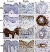
Keratin profiling defines epithelial cells of the von Brunn's nest as transitional. Immunoreactive signals for K13, K14, and K17 are indicated by the brown color. AC: no primary antibody control. DF: K13 antibody treatment. GI: K14 antibody treatment. JL: K17 antibody treatment. A, D, G, and J: human foreskin. B, E, H, and K: human bladder mucosa. C, F, I, and L: representative von Brunn's nest. ED, epidermis; D, dermis; BM, basement membrane; LP, lamina propria; S, superficial layer; I, intermediate layer; B, basal layer; U, urothelium; N, von Brunn's nest; L, lumen of von Brunn's nest. Bar = 20 μm, magnification = ×40.
Fig. 4.
Fig. 4.
K7 is a potential marker of von Brunn's nest maturation in the exstrophic bladder. Shown are immunoreactive signals from a mouse monoclonal IgG specific for K7 (brown). Sections were counterstained with hematoxylin. Shown is immunohistochemistry of the different morphological stages (BD) of Von Brunn's nest in the same tissue section; A is the negative control. BD: expression of K7 as a function of the morphology of von Brunn's nests. Bar = 20 μm, magnification = ×40.
Fig. 5.
Fig. 5.
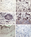
The superficial layer of von Brunn's nest epithelial tissue does not express uroplakin IIIb. Immunoreactive signals for uroplakin IIIb are indicated by the brown color. Specimens were counterstained with hematoxylin (blue). A: superficial layer and lumen of von Brunn's nest. Note the lack of signal in the superficial cell layer. N, von Brunn's nest; S, superficial cell layer. Bar = 10 μm, magnification = ×100. B: normal human bladder positive control specimen. Bar = 10 μm, magnification = ×100. C: normal human ureter positive control specimen. Bar = 20 μm, magnification = ×40. D: normal anterior urethra from adult male specimen. Bar = 20 μm, magnification = ×40.
Fig. 6.
Fig. 6.
von Brunn's nests are absent in normal bladder. Shown are Masson's trichrome-stained sections of formalin-fixed, paraffin-embedded bladder mucosa. A: normal bladder mucosa from 39-wk-old fetus. B: normal bladder mucosa from 2.5-yr female. C: normal bladder mucosa from 7-yr-old female. D: bladder mucosa from 13-yr-old male. LP, lamina propria; URO, urothelium. Bar = 50 μm, magnification = ×20.
Fig. 7.
Fig. 7.
FGF-10 (−/−) mice exhibit an abnormal urothelium but do not develop von Brunn's nests or exstrophy. A: gross anatomy of (−/−) urinary tract. Shown is dissection of (−/−) pup at 19 days postconception (E17.5, Theiler stage 27). B: histology of (+/+) bladder urothelium. Shown is Masson's trichrome stain of wild-type bladder mucosa. U, urothelium; LP, lamina propria; L, lumen; dotted line, basement membrane. C: histology of (−/−) bladder urothelium. Shown is Masson's trichrome stain of bladder mucosa. D and E: systemic administration of FGF-10 rescues abnormal urothelium of FGF-7 (−/−) mice. Bladder was stained with Masson's trichrome: mesenchymal collagen (blue) and epithelium (red). D: daily injection of vehicle for 14 days. Black arrow, abnormal urothelium. E: FGF-10 injection (5 mg/kg) for 14 days. White arrows, responsive basal cells in cell cycle; green arrows, restoration of superficial cell layer.
Fig. 8.
Fig. 8.
Immunoreactive FGF-10 is detected in von Brunn's nests, in cells of the lamina propria, and in the urothelium of the exstrophic bladder. Shown are immunoreactive signals from a mouse monoclonal IgG specific for FGF-10 (brown). Section in A and B are counterstained with hematoxylin. A: no primary antibody control. D: exstrophic bladder urothelium. A, B, and D: bar = 20 μm; magnification = ×40. C: bar = 20 μm, magnification = ×20. N, nest; LP, lamina propria; U, urothelium. Arrowheads, round cell in lamina propria immunopositive for FGF-10. Yellow arrows, fibroblast immunopositive for FGF-10. Yellow K, keratinization.
Fig. 9.
Fig. 9.
FGF-10 receptor is a marker for the earliest stages of von Brunn's nest development in the exstrophic bladder. Shown are immunoreactive signals from a mouse monoclonal IgG specific for the FGF-10 receptor (BD: brown/purple; E and F: green). Sections counterstained with hematoxylin (AD: blue; E and F: gold). A: no primary antibody control. BF: antibody treatment. NE, nest epithelium; NL, nest lumen; LP, lamina propria AC and E: bar = 20 μm, magnification = ×40. D and F: bar = 10 μm, magnification = ×100. Insets in C and E correspond to D and F, respectively. Rp, receptor positive. Arrowheads in D and F, Rp single cell; arrows in D and F, Rp cell cluster. E and F: to better distinguish the hematoxylin blue color with the brown/purple color of the diformazan precipitate (corresponding to receptor signals), raw images of C and D were processed as follows: 1) color intensity and luminance adjusted to 200 with Match Color tool of Photoshop, yielding C and D; 2) resized with bicubic resampling to match the resolution of A and B; 3) image inversion; and 4) color intensity and luminance adjusted to 200, yielding E and F.
Fig. 10.
Fig. 10.
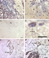
Multiple sites of mRNA synthesis of the FGF-10 receptor in the exstrophic bladder. Shown are in situ hybridization antisense signals (dark brown in AE) derived from an RNA probe complementary for receptor mRNA. A: region of the lamina propria devoid of von Brunn's nests. BD: region of the lamina propria that is rich in von Brunn's nests; arrows point to the clusters formed by FGF-10 receptor mRNA-positive cells; arrowheads point to single positive cells. E: exstrophic bladder urothelium. U, urothelium, LP, lamina propria; arrows point to intense signals at a squamous keratinized epithelial cell layer that lines the bladder lumen (BL). F: sense signals from a region of the lamina propria that is rich in von Brunn's nest. All sections except E were counterstained with hematoxylin. N, nest epithelium. AE: bar = 20 μm, magnification = ×40. F: bar = 10 μm, magnification = ×100.
Fig. 11.
Fig. 11.
Association of FGF-10 receptor mRNA synthesis with blood vessels of the exstrophic bladder. Shown are in situ hybridization signals (brown) derived from annealing of a specific RNA probe to receptor mRNA. All sections except C were counterstained with hematoxylin (blue). A, B, and DF: signals from antisense probe. C: signals from antisense probe. BV, blood vessel; LP, lamina propria; N, nest; arrowheads, positive nucleated blood cells. D: image is a magnified area from the central, positive cells in B. A, B, and D: bar = 20 μm, magnification = ×40. C: bar = 20 μm, magnification = ×20. E and F: bar = 25 μm, magnification = ×40.
Fig. 12.
Fig. 12.
Novel keratin immunostaining patterns are observed in epithelial cells cultured in vitro from the mucosa of the exstrophic bladder. Widefield fluorescence images are shown in A, C, and D. The differential interference contrast image shown in B corresponds to the widefield fluorescence image in C. Shown are immunoreactive profiles for K14 (green signal in A) and K7 (green signal in C and D). F-actin signals are displayed in red in A. DNA signals are displayed in blue in A, C, and D. A, B, and C: magnification = ×40. D: magnification = ×63. Yellow dotted ellipse in D, unique pattern of small K7-positive foci; yellow arrow in C, K7 dome; white arrowheads in C and D, unique pattern of ∼5-μm K7-positive islands. Bar = 20 μm for all images.

Similar articles

Cited by

References

    1. Al Ahmadie H, Gomez AM, Trane N, Bove KE. Giant botryoid fibroepithelial polyp of bladder with myofibroblastic stroma and cystitis cystica et glandularis. Pediatr Dev Pathol 6: 179–181, 2003 - PubMed
    1. Bagai S, Rubio E, Cheng JF, Sweet R, Thomas R, Fuchs E, Grady R, Mitchell M, Bassuk JA. Fibroblast growth factor-10 is a mitogen for urothelial cells. J Biol Chem 277: 23828–23837, 2002 - PubMed
    1. Bassuk JA, Birkebak T, Rothmier JD, Clark JM, Bradshaw A, Muchowski PJ, Howe CC, Clark JI, Sage EH. Disruption of the Sparc locus in mice alters the differentiation of lenticular epithelial cells and leads to cataract formation. Exp Eye Res 68: 321–331, 1999 - PubMed
    1. Bassuk JA, Grady R, Mitchell M. The molecular era of bladder research. Transgenic mice as experimental tools in the study of outlet obstruction. J Urol 164: 170–179, 2000 - PubMed
    1. Beare JB, Tormey AR, Jr, Wattenberg CA. Exstrophy of the urinary bladder complicated by adenocarcinoma. J Urol 76: 583–594, 1956 - PubMed

Publication types

MeSH terms

LinkOut - more resources