. 2010 Oct 22;285(43):33510-33519.
doi: 10.1074/jbc.M110.122093. Epub 2010 Aug 18.

Factors from human embryonic stem cell-derived fibroblast-like cells promote topology-dependent hepatic differentiation in primate embryonic and induced pluripotent stem cells

Affiliations

Factors from human embryonic stem cell-derived fibroblast-like cells promote topology-dependent hepatic differentiation in primate embryonic and induced pluripotent stem cells

Hsiang-Po Huang et al. J Biol Chem. .

Abstract

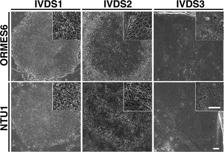
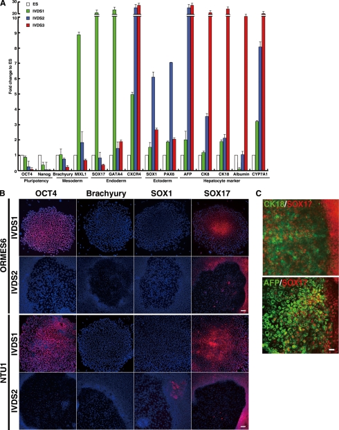
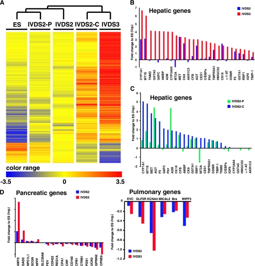
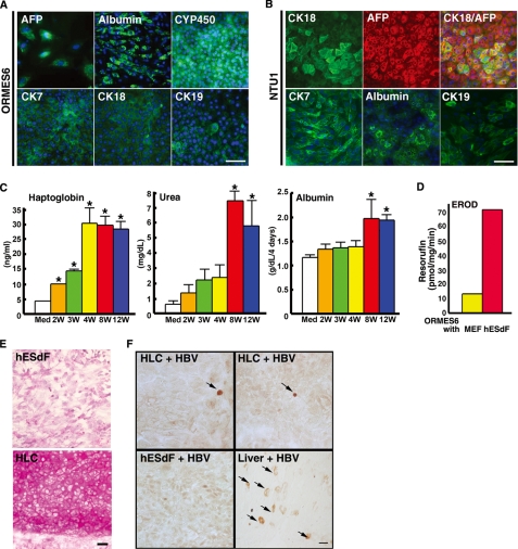
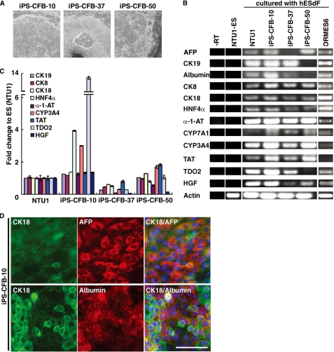
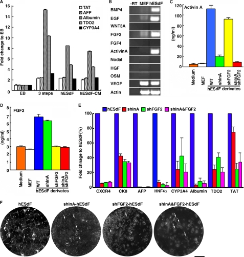
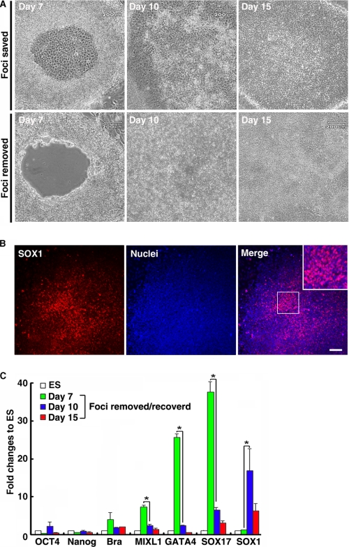
The future clinical use of embryonic stem cell (ESC)-based hepatocyte replacement therapy depends on the development of an efficient procedure for differentiation of hepatocytes from ESCs. Here we report that a high density of human ESC-derived fibroblast-like cells (hESdFs) supported the efficient generation of hepatocyte-like cells with functional and mature hepatic phenotypes from primate ESCs and human induced pluripotent stem cells. Molecular and immunocytochemistry analyses revealed that hESdFs caused a rapid loss of pluripotency and induced a sequential endoderm-to-hepatocyte differentiation in the central area of ESC colonies. Knockdown experiments demonstrated that pluripotent stem cells were directed toward endodermal and hepatic lineages by FGF2 and activin A secreted from hESdFs. Furthermore, we found that the central region of ESC colonies was essential for the hepatic endoderm-specific differentiation, because its removal caused a complete disruption of endodermal differentiation. In conclusion, we describe a novel in vitro differentiation model and show that hESdF-secreted factors act in concert with regional features of ESC colonies to induce robust hepatic endoderm differentiation in primate pluripotent stem cells.


Figures

FIGURE 1.
FIGURE 1.
Stages of hESdF induced regional differentiation in human and monkey ESC colonies. The in vitro differentiation stages (IVDS1, IVDS2, and IVDS3) of ESC colonies were defined by the temporal morphological change of human or monkey ESCs after replating onto hESdF feeders. IVDS1 colonies (days 1–5) resembled ESC colonies; IVDS2 colonies (days 6–10) contained central foci filled with flattened and spiky cells. IVDS3 colonies (days 10–15) contained hepatocyte-like cells. Scale bar, 50 μm.
FIGURE 2.
FIGURE 2.
hESdFs promote a sequential endodermal-to-hepatic differentiation in human and monkey ESCs. A, QRT-PCR analysis of IVDS 1, 2, and 3 for pluripotency-related markers (Oct4 and Nanog), mesodermal markers (Brachyury MIXL1), endodermal markers (SOX17, GATA4, and CXCR4), ectodermal markers (SOX1 and PAX 6), and hepatocyte markers (AFP, CK8, CK18, albumin, and CYP7A1). B, the expression of Oct4 (pluripotency-related), early ectodermal marker SOX1, early mesodermal Brachyury, and early endodermal SOX17 in differentiating human and monkey ESC colonies of IVDS 1 and 2 were analyzed by ICC. C, co-expression of endodermal marker SOX 17 with hepatocyte marker CK18 or AFP is as indicated. IVDS2 foci derived from monkey ESC were stained for SOX17 (red) and CK18 (green) or AFP (green). Scale bar, 25 μm.
FIGURE 3.
FIGURE 3.
Microarray analysis of the spatial and temporal global gene expression pattern in differentiated monkey ES induced by hESdF co-culturing. A, hierarchical clustering of ESCs and cells of IVDS2, IVDS2-P, IVDS2-C, and IVDS3 according to genes that were highly differentially expressed (>10-fold change) between ESCs and IVDS3 differentiated cells as highlighted by heat map. B and C, differential expression (>2-fold change) of genes known to be highly expressed in the liver by UniGene transcriptome cluster annotation were used to analyze the expression biases of cells of IVDS2 and IVDS3 (B) or IVDS2-P and IVDS2-C (C). D, genes known to be highly expressed in the pancreas and lung by UniGene transcriptome cluster annotation were used to analyze the expression biases of cells of IVDS2 and IVDS3 (B).
FIGURE 4.
FIGURE 4.
The HLCs derived from primate ESCs co-cultured with hESdFs exhibited mature and functional phenotypes of hepatocytes. A and B, ICC analysis of hepatocyte marker expression in human (NTU1) and monkey (ORMES-6) ESC-derived HLCs showed that the majority of the HLCs were positively stained by the indicated hepatic markers. The nuclei were stained with DAPI (blue). Scale bar, 25 μm. C, temporal production of haptoglobin, albumin, and urea hepatocyte proteins during differentiation and maturation of monkey ESC-derived HLCs under hESdF co-culture conditions. The production of haptoglobin, albumin, and urea in the medium of differentiated cells increased in parallel with differentiation duration (Med, medium; 2W, 2 weeks; 4W, 4 weeks; 8W, 8 weeks; 12W, 12 weeks). The data correspond to the averages and S.D. of triplicate experiments, except the albumin data, which were the averages of four independent experiments. Significant differences between each time point and media are labeled (*, p < 0.05). D, ethoxyresorufin-O-deethylase assay showed that differentiated cells had a higher activity than control cells. E, periodic acid-Schiff staining showed glycogen storage in HLCs (lower panel), whereas the hESdF feeder cells were negative. F, immunoperoxidase staining showed positive nuclear staining of HBcAg (arrows, upper panels) in monkey ESC-derived HLCs infected with serum from patients with HBV infection for 3 days. The feeder cells (left lower panel) were negative. An HBV-infected liver section (right lower panel), stained for both cytoplasmic and nuclear (arrows); HBcAg was the positive control. Scale bar, 25 μm.
FIGURE 5.
FIGURE 5.
Derivation of human iPSCs and characterization of iPSC-derived HLCs under hESdF co-culture conditions. A, phase contrast images of three human iPSC lines (cell lines 10, 37, and 50), which showed characteristic hepatocyte-like morphology after 3 weeks co-culture with feeder hESdFs. B, RT-PCR analysis of hepatic markers (CK8, CK18, CK19, AFP, Albumin, HNF4α, α-1-AT, CYP3A4, CYP7A1, TAT, TDO2, and HGF) expression in the HLCs derived from human ESCs (NTU1) and monkey ESCs (ORMES6) and three different iPSC clones, and in control ESCs (NTU1) without differentiation. C, QRT-PCR analysis of total RNA isolated from HLCs derived from iPSC-CFB-10, -37, and -50 clones for CK8, CK18, CK19, HNF4α, α-1-AT, CYP3A4, TAT, TDO2, and HGF expression. For each sample, relative expression levels were normalized to corresponding levels in human ESCs (NTU1). The data correspond to the averages and standard deviations of triplicate experiments. D, ICC analysis of hepatocyte markers in human iPSC-derived HLCs. HLCs derived from iPSC-CFB-10 clone at day 15 of differentiation were double-stained with anti-human CK18 (green) and AFP (red) or albumin (red) as indicated. The nuclei were stained with DAPI (blue). Scale bar, 25 μm.
FIGURE 6.
FIGURE 6.
hESdF secreted factors promoting hepatic differentiation in pluripotent stem cells. A, QRT-PCR analysis of hepatic markers TAT, AFP, albumin, TDO2, and CYP3A4 in ESCs cultured under EB-based differentiation conditions (29), three-step culture conditions, and hESdF co-culture conditions and in hESdF-CM (27). The cells were harvested for analysis 10 days post-differentiation. Gene expression levels are presented as the relative change of the 2−ΔΔCt value relative to that of day 10 EB-based differentiation conditions. The data correspond to the averages and standard deviations of triplicate experiments. B, RT-PCR analysis of BMP4, EGF, FGF2, WNT3A, FGF2, FGF4, activin A, Nodal, HGF, oncostatin M (OSM), and VEGF in hESdFs. Actin was used as a loading control, and a reaction without reverse transcriptase (−RT) was applied to rule out genomic DNA contamination. C and D, ELISAs for activin A (C) and FGF2 protein (D) expression in conditioned media derived from MEFs, hESdFs, and hESdFs transduced with short hairpin RNA specific to INHBA (shInA) and/or FGF2 (shFGF2). E, QRT-PCR analysis for hepatic markers in monkey ESCs co-cultured with activin A and/or FGF2 knockdown hESdFs for 15 days. Gene expression levels relative to those in hESdF co-cultured cells are shown. F, morphology of monkey ESC colonies co-cultured with hESdFs and shInA and/or shFGF2 hESdFs for 15 days. The number and size of hepatic foci were dramatically reduced in shFGF2 and/or shInA hESdF group(s). Small hepatic foci (arrows) were occasionally found in the ESC colonies cultured in the modified hESdFs.
FIGURE 7.
FIGURE 7.
Removal of the central foci of monkey ESC colonies from IVDS2 disrupted hepatic endoderm differentiation under hESdF-primed culture conditions. A, the differentiated cells in the central area of ESC colonies at early IVDS2 (day 7) were excised and removed with a glass micropipette. The central area of an ESC colony was repopulated by cells grown from peripheral areas 3 days (day 10) after cell removal. B, ICC for SOX1 on ESC colonies 3 days after cell removal, showing that the central area of ESC colonies was repopulated with SOX1-expressing cells. Scale bar, 25 μm. C, QRT-PCR analysis of total RNA isolated from foci removed IVDS2 (day 7), regenerated cells at 3 (day 10) and 8 days (day 15) after central area removal using primers specific for Oct4, Nanog, Brachyury, MIXL1, GATA4, SOX17, and SOX1. The data correspond to the means and standard deviations of triplicate experiments. *, p < 0.05 by Student's t test.

Similar articles

Cited by

References

    1. Akhter J., Johnson L. A., Gunasegaram A., Riordan S. M., Morris D. L. (2007) Surgeon 5, 155–164 - PubMed
    1. Thomson J. A., Kalishman J., Golos T. G., Durning M., Harris C. P., Becker R. A., Hearn J. P. (1995) Proc. Natl. Acad. Sci. U.S.A. 92, 7844–7848 - PMC - PubMed
    1. Mitalipov S., Kuo H. C., Byrne J., Clepper L., Meisner L., Johnson J., Zeier R., Wolf D. (2006) Stem Cells 24, 2177–2186 - PubMed
    1. Suemori H., Tada T., Torii R., Hosoi Y., Kobayashi K., Imahie H., Kondo Y., Iritani A., Nakatsuji N. (2001) Dev. Dyn. 222, 273–279 - PubMed
    1. Reubinoff B. E., Pera M. F., Fong C. Y., Trounson A., Bongso A. (2000) Nat. Biotechnol. 18, 399–404 - PubMed

Publication types