. 2010 Oct 29;285(44):34202-12.
doi: 10.1074/jbc.M110.134643. Epub 2010 Aug 18.

A 24-residue peptide (p5), derived from p35, the Cdk5 neuronal activator, specifically inhibits Cdk5-p25 hyperactivity and tau hyperphosphorylation

Affiliations

A 24-residue peptide (p5), derived from p35, the Cdk5 neuronal activator, specifically inhibits Cdk5-p25 hyperactivity and tau hyperphosphorylation

Ya-Li Zheng et al. J Biol Chem. .

Abstract

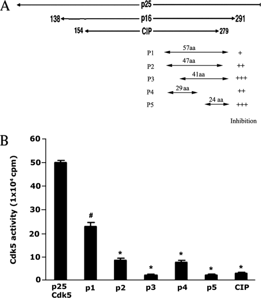
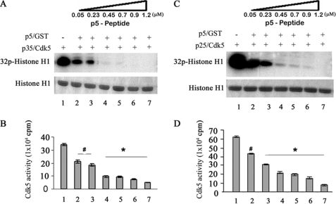
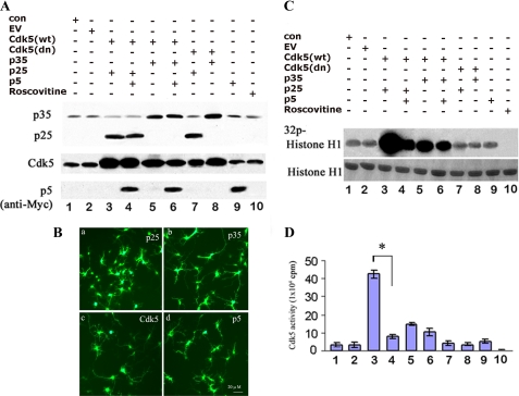
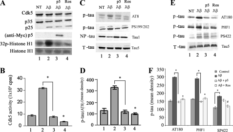
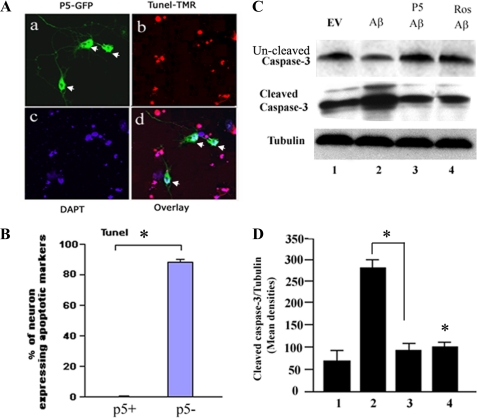
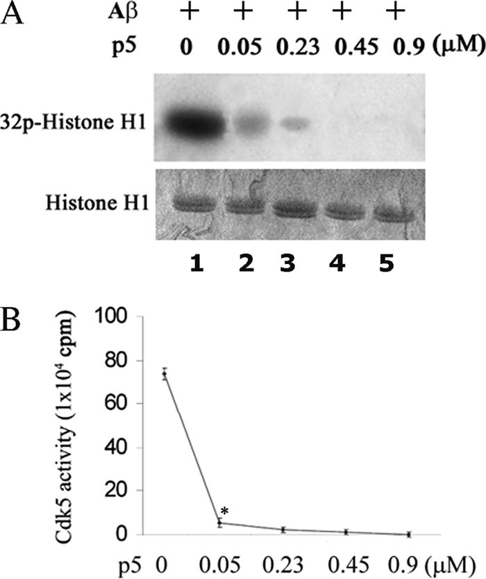
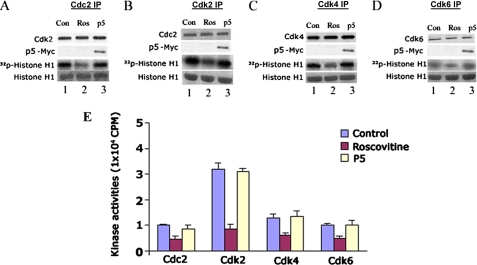
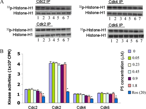
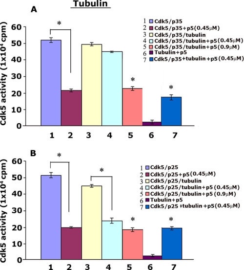
The activity of Cdk5-p35 is tightly regulated in the developing and mature nervous system. Stress-induced cleavage of the activator p35 to p25 and a p10 N-terminal domain induces deregulated Cdk5 hyperactivity and perikaryal aggregations of hyperphosphorylated Tau and neurofilaments, pathogenic hallmarks in neurodegenerative diseases, such as Alzheimer disease and amyotrophic lateral sclerosis, respectively. Previously, we identified a 125-residue truncated fragment of p35 called CIP that effectively and specifically inhibited Cdk5-p25 activity and Tau hyperphosphorylation induced by Aβ peptides in vitro, in HEK293 cells, and in neuronal cells. Although these results offer a possible therapeutic approach to those neurodegenerative diseases assumed to derive from Cdk5-p25 hyperactivity and/or Aβ induced pathology, CIP is too large for successful therapeutic regimens. To identify a smaller, more effective peptide, in this study we prepared a 24-residue peptide, p5, spanning CIP residues Lys(245)-Ala(277). p5 more effectively inhibited Cdk5-p25 activity than did CIP in vitro. In neuron cells, p5 inhibited deregulated Cdk5-p25 activity but had no effect on the activity of endogenous Cdk5-p35 or on any related endogenous cyclin-dependent kinases in HEK293 cells. Specificity of p5 inhibition in cortical neurons may depend on the p10 domain in p35, which is absent in p25. Furthermore, we have demonstrated that p5 reduced Aβ(1-42)-induced Tau hyperphosphorylation and apoptosis in cortical neurons. These results suggest that p5 peptide may be a unique and useful candidate for therapeutic studies of certain neurodegenerative diseases.


Figures

FIGURE 1.
FIGURE 1.
Identification of Cdk5-inhibitory peptides derived from p25. A, a schematic diagram of p25 truncated peptides showing the CIP peptide from which five short peptides (p1–p5) were derived. p5 was the shortest and best inhibitor of Cdk5-p25 in an in vitro assay with histone H1 as substrate. B, a histogram summary comparison of the inhibitory effects of peptides derived from CIP in a standard in vitro pad assay of Cdk5-p25 activity. p3 and p5 were the most effective inhibitors under these conditions, reducing activity more than 90%. Data represent the mean ± S.E. (error bars) of three independent experiments. *, p < 0.01. aa, amino acids.
FIGURE 2.
FIGURE 2.
p5 equally inhibits both Cdk5-p25 and Cdk5-p35 activities in vitro (a dose-dependent analysis). Activated Cdk5-p35 and Cdk5-p25 were used in a comparative test tube assay of the inhibitory effect of p5 with histone H1 as the substrate. p5 as p5-GST was added at a range of concentrations from 0.05 to 1.2 μm. After SDS-PAGE, autoradiographs were prepared as shown in A and C, and pad assay scintillation counts were quantified from three separate experiments as summarized in the bar graphs (B and D). Note that initially, the Cdk5-p25 activity is twice that of the Cdk5-p35 activity. The levels of inhibition, however, are almost identical, although it might appear from the autoradiograms that the Cdk5-p35 activity is more sensitive because it seems completely inhibited at 0.45 μm, whereas Cdk5-p25 activity is evident at least to 0.9 μm in the sample shown. This is due to the initially higher activity of the Cdk5-p25 complex. The quantitative data (B and D), however, show that p5 equally inhibits each Cdk5 complex in a dose-dependent manner when compared with lanes 1 in B and D, respectively; at 0.05 μm (lane 2), p5 peptide inhibits 36% of Cdk5-p35 activity and ∼33% of Cdk5-p25 activity, whereas 0.7 μm (lane 5) inhibits both complexes ∼70%, and finally at 1.2 μm (lane 6), both are inhibited ∼90%. *, p < 0.01; #, p < 0.05. Error bars, S.E.
FIGURE 3.
FIGURE 3.
p5 inhibits Cdk5-p25 activity without affecting endogenous Cdk5-p35 activity in cortical neurons. A, to determine the effect of p5 on Cdk5-p35 and Cdk5-p25 activity in infected neurons, E-18 rat cortical neurons after 5 DIC were infected with different gene constructs using adenoviral vector as follows: co-infected with Cdk5-p25, Cdk5-p35, dominant negative Cdk5-p25, and dominant negative Cdk5-p35 or triply infected with GFP-Myc-p5, except for cells that were infected with GFP-Myc-p5 alone and one group of uninfected cells treated with 20 μm roscovitine as a control. After SDS-PAGE and Western analysis with the specific antibodies, the expression level of each of the constructs is shown. B, the expressions of cells infected with p25 (a), p35 (b), Cdk5 (c), and p5 (d). C, Cdk5 activity was measured in Cdk5 immunoprecipitates (IP) from lysates of these infected cells (from A) using histone H1 as a substrate. The top panel is the autoradiograph, and the bottom panel shows the corresponding Coomassie-stained histone H1 bands. D, a histogram showing quantitative scintillation count data (cpm) from corresponding pad assays of kinase activities from the same lysates. Data represent means ± S.E. (error bars) of three separate experiments. *, p < 0.01.
FIGURE 4.
FIGURE 4.
Aβ-mediated Cdk5-deregulated phosphorylation of Tau is inhibited by p5 in cortical neurons. 7 DIC cortical neurons from E-18 rat brain were infected with p5 (Myc-p5) and treated with 10 μm Aβ(1–42) for 6 h, lysed, and prepared for Cdk5 immunoprecipitation, Western blot analysis, and kinase assays. A, Western blots show the formation of p25 from p35 as a result of the stress induced by Aβ (lanes 2–4), accompanied by a significant increase in histone phosphorylation (compare lane 2 with lane 1). In the presence of p5, however, the activity is decreased by 70% to control levels, although p25 is still present (compare lane 3 with lane 2). Neurons treated with 20 μm roscovitine show almost complete inhibition (lane 4). B, the scintillation count results of three separate pad assay experiments of the above lysates are plotted in the bar graph as means ± S.E. (error bars). C, Western blots of these same Aβ-treated cells infected with and without p5 exhibit Aβ-induced Tau phosphorylation at PHF Tau sites, Ser202, Thr205, and Ser199, as detected by AT8 antibody (compare lanes 1 and 2). Cells infected with p5, however, show base-line levels of Tau phosphorylation comparable with that produced by roscovitine (lanes 3 and 4), indicating p5 rescue from Aβ-induced Tau hyperphosphorylation. Another Tau antibody specific for sites Ser199/Ser202 is also shown, confirming the above result. Tau1 recognizes nonphosphorylated Tau, whereas Tau5 identifies total Tau, phosphorylated as well as non-phosphorylated. D, a histogram of optical density data of phosphorylation by AT8 was prepared from three separate experiments. E, to show that several other phosphorylated Tau sites were rescued by p5, Western blots of similarly treated lysates were immunoreacted with PHF antibodies AT180 (Thr(P)231), Ser(P)404, and Ser(P)422. Comparison of lanes 1 and 2 shows significantly elevated Aβ-induced phosphorylation at sites Thr231 and Ser404, with lower levels at Ser422. The addition of p5 reduces phosphorylation at Thr231 and Ser404 with only a modest effect on Ser422 (compare lanes 2 and 3). F, data from three separate experiments are quantified in the bar graph. *, p < 0.01; #, p < 0.05.
FIGURE 5.
FIGURE 5.
Aβ-induced apoptosis is prevented in the presence of p5. A, E-18 cortical neurons grown for 3 days were transfected with or without GFP-p5. After 24 h, they were treated with Aβ for 6 h, fixed, and prepared for a TUNEL apoptosis assay. It can be seen in the fluorescence images that p5-transfected neurons expressing GFP (a) are prevented from undergoing apoptosis as seen in the overlay in d. B, The data are quantified in the bar graph showing that virtually all p5-transfected cells were protected from Aβ-induced apoptosis. The percentage of GFP- and TUNEL-expressing neurons was determined by counting at least 100 DAPI-stained nuclei in each of five fields. Data from three experiments are shown. C, the cortical neurons infected with or without p5 were treated with Aβ for 6 h, lysed, and prepared for Western blots using caspase-3 and cleaved caspase-3 antibody expression as a measure of apoptosis. Cells infected with p5 show a marked reduction in cleaved caspase-3 expression compared with Aβ-treated cells without p5 (compare lanes 2 and 3) (panel 2) to a level comparable with the effect of roscovitine (lane 4). The uncleaved caspase-3 is lower in Aβ-induced cells (panel 1, lane 2) while higher in cells treated with p5 or roscovitine. D, the data from three experiments are expressed in a bar graph as means ± S.E. (error bars). *, p < 0.01.
FIGURE 6.
FIGURE 6.
p5 at low concentrations inhibits Cdk5-p25 activity induced by Aβ in cortical neurons. A, 7 DIC cortical neurons were treated with 10 μm Aβ(1–42) or PBS for 6 h, and the harvested cells were lysed for Cdk5 immunoprecipitation using anti-Cdk5 antibody. Immunoprecipitates were then used as an enzyme in in vitro kinase assays with increasing concentrations of p5 peptide. The top panel is the autoradiograph, and the bottom panel shows the corresponding Coomassie-stained histone H1 bands. High levels of induced activity are rapidly inhibited by the addition of p5. B, a line graph shows that Cdk5-p25 activities induced by Aβ were inhibited by p5 in a dose-dependent manner with most of the inhibition (90%) exhibited at 0.05 μm, the lowest concentration. Data represent means ± S.E. from three experiments. *, p < 0.01.
FIGURE 7.
FIGURE 7.
p5 does not inhibit activity of cell cycle Cdks. To study the effect of p5 on cycling cells, a group of proliferating HEK293 cells were infected with p5-Myc, the second group were infected with EV as control, and the third group were infected with EV but treated with 20 μm roscovitine. Cell lysates from each group were immunoprecipitated with specific antibodies to Cdc2 (A), Cdk2 (B), Ckd4 (C), and Cdk6 (D), respectively. The immunoprecipitates were used for Western blotting and kinase assays with histone H1 as substrate. The expression of p5-Myc, the specific kinases as seen in blots (lanes 1 and 2), and the phosphorylating activities (presented as radioautographs, lane 3) are shown in A–D. The scintillation count data (means ± S.E. (error bars)) from three pad assay experiments in each case are quantified in the bar graphs (E). It is clear in each case that roscovitine inhibits ∼50% of endogenous Cdk activity (compare bars 2 with bars 1), whereas p5 has virtually no effect, as evident from the bar graph, (comparing bars 3 with bars 1).
FIGURE 8.
FIGURE 8.
High concentrations of p5 do not inhibit activities of cell cycle Cdks. A dose-dependent analysis shows the effect of p5 on Cdk activities in vitro. Non-treated and non-transfected HEK293 cell lysates were subjected to immunoprecipitation using anti-Cdc2, anti-Cdk2, anti-Cdk4, and anti-Cdk6 antibodies. The immunoprecipitates were used for standard kinase pad assays in the presence of increasing concentrations of p5 peptide (0.05–1.8 μm). A, the phosphorylating activities at different concentrations of p5 (lanes 1–6) and 20 μm roscovitine (positive control, lane 7) for each Cdk are shown in radioautographs. Except for roscovitine, activities in the presence of p5 are relatively unaffected. B, a bar graph summarizes scintillation counting data from three experiments (means ± S.E.) of histone H1 phosphorylation. Note that only roscovitine has a significant effect on Cdk endogenous activities (∼50%; compare lane 7 with lane 1), whereas p5, even at the highest concentration of 1.8 μm, exhibits virtually no inhibition. *, p < 0.01.
FIGURE 9.
FIGURE 9.
Polymerized tubulin protects Cdk5-p35 activity from p5 inhibition in vitro. Active Cdk5-p35 and Cdk5-p25 were preincubated with tubulin in PEM buffer supplemented with 1 mm GTP at 35 °C for 45 min to 1 h (under conditions to promote polymerization). p5 at 0.45 or 0.9 μm was added, and the mixture was then subjected to in vitro kinase assay. Quantification of Cdk5-p35 (A) and Cdk5-p25 (B) activities showed that p5 inhibits preincubated Cdk5-p25 activity ∼50% (B, compare 3 and 4) but has no effect on preincubated Cdk5-p35 activity (A, compare 3 and 4). At a higher concentration of p5 (0.9 μm), both kinase complexes are inhibited equally (compare 5 in A and B). The importance of polymerization during preincubation is seen in lane 7 of both A and B; in the presence of unpolymerized tubulin, p5 inhibits both kinases equally. The concentrations of Cdk5-p35 and Cdk5-p25 were matched to exhibit equal activity. Data represent mean ± S.E. of three experiments. *, p < 0.01.

Similar articles

Cited by

References

    1. Chae T., Kwon Y. T., Bronson R., Dikkes P., Li E., Tsai L. H. (1997) Neuron 18, 29–42 - PubMed
    1. Cheung Z. H., Fu A. K., Ip N. Y. (2006) Neuron 50, 13–18 - PubMed
    1. Dhavan R., Tsai L. H. (2001) Nat. Rev. Mol. Cell Biol. 2, 749–759 - PubMed
    1. Gilmore E. C., Ohshima T., Goffinet A. M., Kulkarni A. B., Herrup K. (1998) J. Neurosci. 18, 6370–6377 - PMC - PubMed
    1. Lai K. O., Ip N. Y. (2009) Biochim. Biophys. Acta 1792, 741–745 - PubMed

Publication types