. 2010 Oct 22;285(43):33520-33529.
doi: 10.1074/jbc.M110.158378. Epub 2010 Aug 18.

γ-Tocotrienol but not γ-tocopherol blocks STAT3 cell signaling pathway through induction of protein-tyrosine phosphatase SHP-1 and sensitizes tumor cells to chemotherapeutic agents

Affiliations

γ-Tocotrienol but not γ-tocopherol blocks STAT3 cell signaling pathway through induction of protein-tyrosine phosphatase SHP-1 and sensitizes tumor cells to chemotherapeutic agents

Ramaswamy Kannappan et al. J Biol Chem. .

Retraction in

Abstract

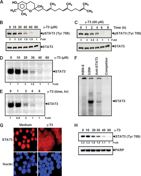
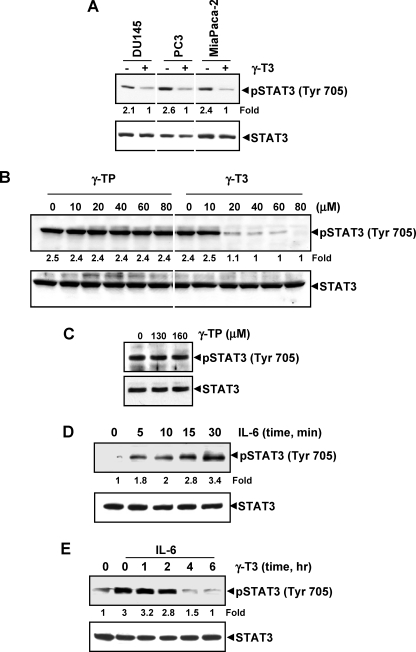
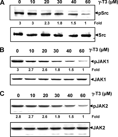
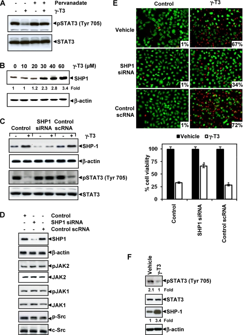
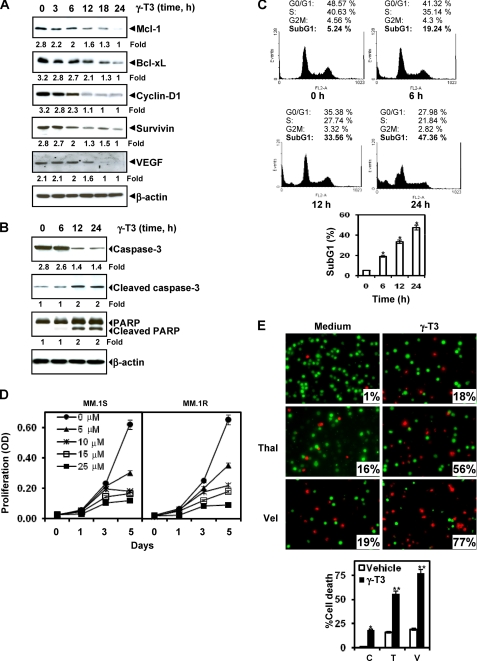
Although γ-tocotrienol (T3), a vitamin E isolated primarily from palm and rice bran oil, has been linked with anticancer activities, the mechanism of this action is poorly understood. In this study, we investigated whether γ-T3 can modulate the STAT3 cell signaling pathway, closely linked to inflammation and tumorigenesis. We found that γ-T3 but not γ-tocopherol, the most common saturated form of vitamin E, inhibited constitutive activation of STAT3 in a dose- and time-dependent manner, and this inhibition was not cell type-specific. γ-T3 also inhibited STAT3 DNA binding. This correlated with inhibition of Src kinase and JAK1 and JAK2 kinases. Pervanadate reversed the γ-T3-induced down-regulation of STAT3 activation, suggesting the involvement of a protein-tyrosine phosphatase. When examined further, we found that γ-T3 induced the expression of the tyrosine phosphatase SHP-1, and gene silencing of the SHP-1 by small interfering RNA abolished the ability of γ-T3 to inhibit STAT3 activation, suggesting a vital role for SHP-1 in the action of γ-T3. Also γ-T3 down-modulated activation of STAT3 and induced SHP-1 in vivo. Eventually, γ-T3 down-regulated the expression of STAT3-regulated antiapoptotic (Bcl-2, Bcl-xL, and Mcl-1), proliferative (cyclin D1), and angiogenic (VEGF) gene products; and this correlated with suppression of proliferation, the accumulation of cells in sub-G(1) phase of the cell cycle, and induction of apoptosis. This vitamin also sensitized the tumor cells to the apoptotic effects of thalidomide and bortezomib. Overall, our results suggest that γ-T3 is a novel blocker of STAT3 activation pathway both in vitro and in vivo and thus may have potential in prevention and treatment of cancers.


Figures

FIGURE 1.
FIGURE 1.
γ-T3 inhibits constitutively active STAT3 in U266 cells. A, chemical structure of γ-T3. B, γ-T3 suppresses phospho-STAT3 levels in a dose-dependent manner. U266 cells (2 × 10⁶/ml) were treated with the indicated concentrations of γ-T3 for 6 h, after which whole-cell extracts were prepared, and 40 μg of protein were resolved on 7.5% SDS-polyacrylamide gel, electrotransferred onto nitrocellulose membrane, and probed for phospho-STAT3. C, γ-T3 suppresses phospho-STAT3 levels in a time-dependent manner. U266 cells (2 × 10⁶/ml) were treated with the 60 μm γ-T3 for the indicated time points, after which Western blotting was done as described above. D, γ-T3 suppresses STAT3 DNA binding in a dose-dependent manner. U266 cells (2 × 10⁶/ml) were treated with the indicated concentrations of γ-T3 for 6 h and analyzed for nuclear STAT3 levels by EMSA. E, U266 cells (2 × 10⁶/ml) were treated with 60 μm γ-T3 for the indicated durations and analyzed for nuclear STAT3 levels by EMSA. F, nuclear extracts from U266 cells were incubated with STAT3 antibody and an unlabeled STAT3 oligonucleotide probe. Nuclear extracts from myeloid leukemia (KBM-5) cells were taken alone. They were then assayed for STAT3 DNA binding by electrophoretic mobility shift assay. G, γ-T3 inhibits translocation of STAT3 to the nucleus. U266 cells (1 × 10⁵/ml) were incubated with or without 60 μm γ-T3 for 6 h and then analyzed for the intracellular distribution of STAT3 by immunocytochemistry. The same slides were counterstained for nuclei with Hoechst (50 ng/ml) for 5 min. H, U266 cells (2 × 10⁶/ml) were treated with the indicated concentrations of γ-T3 for 6 h and analyzed for nuclear STAT3 levels by Western blot.
FIGURE 2.
FIGURE 2.
γ-T3 down-regulates IL-6-induced phospho-STAT3. A, γ-T3 suppresses phospho-STAT3 levels in various cells. DU-145, PC3, and MiaPaCa-2 cells (1 × 10⁶/ml) were treated with 60 μm γ-T3 for 6 h, after which whole-cell extracts were prepared, and protein was resolved on 7.5% SDS-polyacrylamide gel, electrotransferred onto nitrocellulose membrane, and probed for phospho-STAT3. B and C, γ-TP has no effect on activation of STAT3. U266 cells (2 × 10⁶/ml) were treated with the indicated concentrations of either γ-TP or γ-T3 for 6 h, after which the whole-cell extracts were prepared, and phospho-STAT3 levels were detected by Western blot. The same blots were stripped and reprobed with STAT3 antibody to verify equal protein loading. D, γ-T3 suppresses IL-6-induced phospho-STAT3 levels in a dose-dependent manner. MM.1s cells (2 × 10⁶) were treated with IL-6 (10 ng/ml) for the indicated times. Whole-cell extracts were prepared, and phospho-STAT3 level was detected by Western blot. E, γ-T3 suppresses IL-6-induced phospho-STAT3 levels in a time-dependent manner. MM.1s cells (2 × 10⁶) were treated with 60 μm γ-T3 for the indicated times and then stimulated with IL-6 (10 ng/ml) for 30 min. Whole-cell extracts were then prepared and analyzed for phospho-STAT3 by Western blotting.
FIGURE 3.
FIGURE 3.
γ-T3 down-regulates constitutively active Src, Jak1, and Jak2. A, γ-T3 suppresses phospho-Src levels in a dose-dependent manner. U266 cells (2 × 10⁶/ml) were treated with indicated doses of γ-T3 for 6 h, after which whole-cell extracts were prepared, and 40 μg of those extracts were resolved on 10% SDS-PAGE, electrotransferred onto nitrocellulose membranes, and probed with phospho-Src antibody. The same blots were stripped and reprobed with Src antibody to verify equal protein loading. B, γ-T3 suppresses phospho-JAK1 expression in a dose-dependent manner. U266 cells (2 × 10⁶/ml) were treated with indicated doses of γ-T3 for 6 h, after which whole-cell extracts were prepared, and 40 μg of those extracts were resolved on 7.5% SDS-PAGE, electrotransferred onto nitrocellulose membranes, and probed with phospho-JAK1. C, γ-T3 suppresses phospho-JAK2 expression in a dose-dependent manner. U266 cells (2 × 10⁶/ml) were treated with indicated doses of γ-T3 for 6 h, after which whole-cell extracts were prepared, and 40 μg of those extracts were resolved on 7.5% SDS-PAGE, electrotransferred onto nitrocellulose membranes, and probed with phospho-JAK2. The same blots were stripped and reprobed with respective nonphosphorylated protein antibodies to verify equal protein loading.
FIGURE 4.
FIGURE 4.
γ-T3 regulates STAT3 through induction of SHP-1. A, pervanadate reverses the phospho-STAT3 inhibitory effect of γ-T3. U266 cells (2 × 10⁶/ml) were treated with pervanadate (50 μm) and 60 μm γ-T3 for 4 h, after which Western blotting was done as described above. The same blots were stripped and reprobed with STAT3 antibody to verify equal protein loading. B, γ-T3 induces the expression of SHP-1 protein in U266 cells. U266 cells (2 × 10⁶/ml) were treated with indicated concentrations of γ-T3 for 6 h, after which whole-cell extracts were prepared, and 40-μg portions of those extracts were resolved on 10% SDS-PAGE, electrotransferred onto nitrocellulose membranes, and probed with SHP-1 antibody. The same blots were stripped and reprobed with β-actin antibody to verify equal protein loading. C, effect of SHP-1 knockdown on γ-T3-induced expression of SHP-1. SCC4 cells (1 × 10⁵/ml) were transfected with either scrambled or SHP-1-specific siRNA (50 nm). After 48 h, cells were treated with 60 μm γ-T3 for 6 h, and whole-cell extracts were subjected to Western blot analysis for SHP-1, p-STAT3, and STAT3. D, effect of SHP-1 knockdown on the expression levels of constitutively active JAK1, JAK2, and Src. SCC4 cells (1 × 10⁵/ml) were transfected with either scrambled or SHP-1-specific siRNA (50 nm). After 48 h, cells were treated with 60 μm γ-T3 for 6 h, and whole-cell extracts were subjected to Western blot analysis. E, knockdown of SHP-1 inhibits the apoptotic effect of γ-T3. SCC4 cells (1 × 10⁵/ml) were transfected with either scrambled or SHP-1-specific siRNA (50 nm). After 48 h, cells were treated with 25 μm γ-T3 for 24 h, and the percentage of apoptosis was analyzed by the Live/Dead assay. F, human pancreatic tissues from animals treated with vehicle and γ-T3 (400 mg/kg body weight) for 4 weeks were homogenized and analyzed by Western blot for p-STAT3, STAT3, and SHP-1 expression levels as indicated under “Experimental Procedures.” The blots were stripped nd reprobed with β-actin antibody to verify equal protein loading. *, p < 0.01.
FIGURE 5.
FIGURE 5.
γ-T3 suppresses STAT3-regulated antiapoptotic gene products, induces apoptosis, and potentiates chemotherapeutic agents. A, γ-T3 suppresses STAT3-regulated antiapoptotic gene products. U266 cells (2 × 10⁶/ml) were treated with 25 μm γ-T3 for the indicated time intervals, after which whole-cell extracts were prepared, and 40-μg portions of those extracts were resolved on 10% SDS-PAGE; the membrane was sliced according to molecular weight, and the gel was probed using antibodies against cyclin D1, Bcl-2, Bcl-xL, survivin, and VEGF. The same blots were stripped and reprobed with β-actin antibody to verify equal protein loading. B, γ-T3 induces caspase-3-dependent PARP cleavage. U266 cells were treated with 60 μm γ-T3 for the indicated times, and whole-cell extracts were prepared, separated on SDS-PAGE, and subjected to Western blotting against caspase-3 antibody and PARP antibody. The same blots were stripped and reprobed with β-actin antibody to show equal protein loading. C, γ-T3 causes significant accumulation of cells in the sub-G1 phase. U266 cells (2 × 10⁶/ml) were synchronized by incubation overnight in the absence of serum and then treated with 25 μm γ-T3 for the indicated times, after which the cells were washed, fixed, stained with propidium iodide, and analyzed for DNA content by flow cytometry. D, effects of γ-T3 on the proliferation of multiple myeloma cells. MM cells were plated in triplicate, treated with the indicated concentrations of γ-T3 for the indicated days, and then subjected to 3-(4,5-dimethylthiazol-2-yl)-2,5-diphenyl-2H-tetrazolium bromide (MTT) assay. E, γ-T3 potentiates the apoptotic effect of thalidomide (Thal) and Velcade (Val). U266 cells (1 × 10⁶/ml) were treated with 25 μm γ-T3 and 10 ng/ml thalidomide or 20 nm bortezomib alone or in combination for 24 h at 37 °C. Cells were stained with a Live/Dead assay reagent for 30 min, then analyzed under a fluorescence microscope, and 20 random fields were counted. C, Control; T, Thalidomide; V, Velcade; *, p < 0.01; **; p < 0.05 versus control.
FIGURE 6.
FIGURE 6.
Schematic diagram showing the effect of γ-T3 on STAT3 signaling pathway and apoptosis.

Similar articles

Cited by

References

    1. Sen C. K., Khanna S., Rink C., Roy S. (2007) Vitam. Horm. 76, 203–261 - PMC - PubMed
    1. Sen C. K., Khanna S., Roy S. (2007) Mol. Aspects Med. 28, 692–728 - PMC - PubMed
    1. Sylvester P. W., Shah S. J. (2005) Front. Biosci. 10, 699–709 - PubMed
    1. Yu W., Simmons-Menchaca M., Gapor A., Sanders B. G., Kline K. (1999) Nutr. Cancer 33, 26–32 - PubMed
    1. Ahmad N. S., Khalid B. A., Luke D. A., Ima Nirwana S. (2005) Clin. Exp. Pharmacol. Physiol. 32, 761–770 - PubMed

Publication types

MeSH terms

Substances