. 2010 Sep;256(3):714-23.
doi: 10.1148/radiol.10092311.

Contrast-enhanced dedicated breast CT: initial clinical experience

Affiliations

Contrast-enhanced dedicated breast CT: initial clinical experience

Nicolas D Prionas et al. Radiology. 2010 Sep.

Abstract

Purpose: To quantify contrast material enhancement of breast lesions scanned with dedicated breast computed tomography (CT) and to compare their conspicuity with that at unenhanced breast CT and mammography.

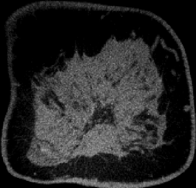
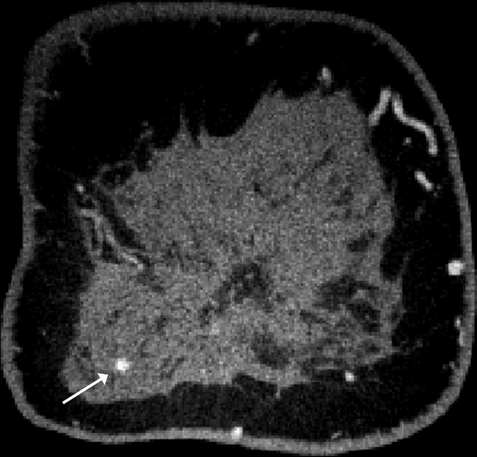
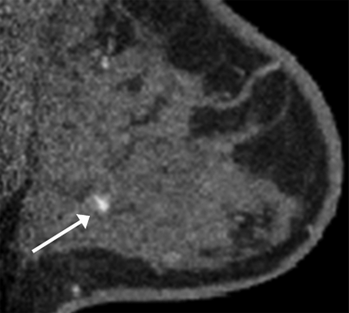
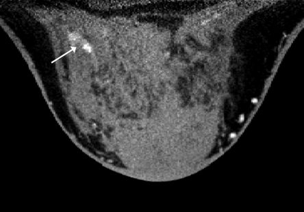
Materials and methods: Approval of the institutional review board and the Radiation Use Committee and written informed consent were obtained for this HIPAA-compliant study. Between September 2006 and April 2009, 46 women (mean age, 53.2 years; age range, 35-72 years) with Breast Imaging Reporting and Data System category 4 or 5 lesions underwent unenhanced breast CT and contrast material-enhanced breast CT before biopsy. Two radiologists independently scored lesion conspicuity for contrast-enhanced breast CT versus mammography and for contrast-enhanced breast CT versus unenhanced breast CT. Mean lesion voxel intensity was measured in Hounsfield units and normalized to adipose tissue intensity on manually segmented images obtained before and after administration of contrast material. Regression models focused on conspicuity and quantified enhancement were used to estimate the effect of pathologic diagnosis (benign vs malignant), lesion type (mass vs calcifications), breast density, and interradiologist variability.

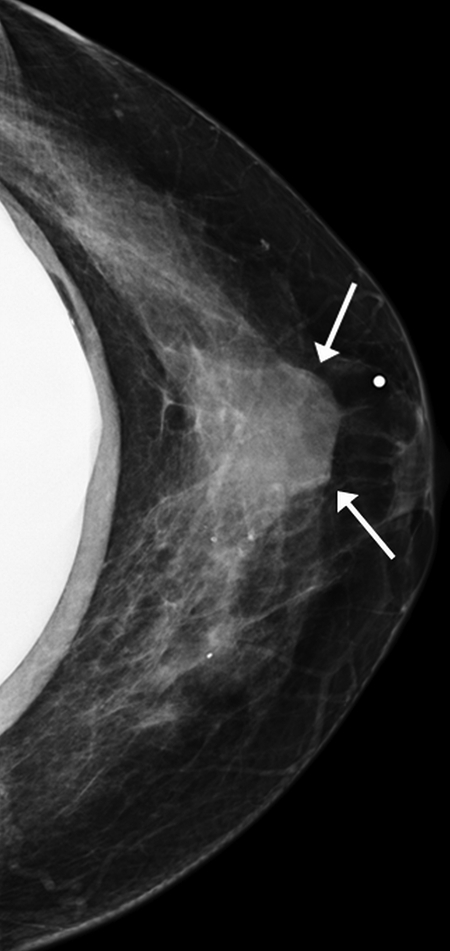
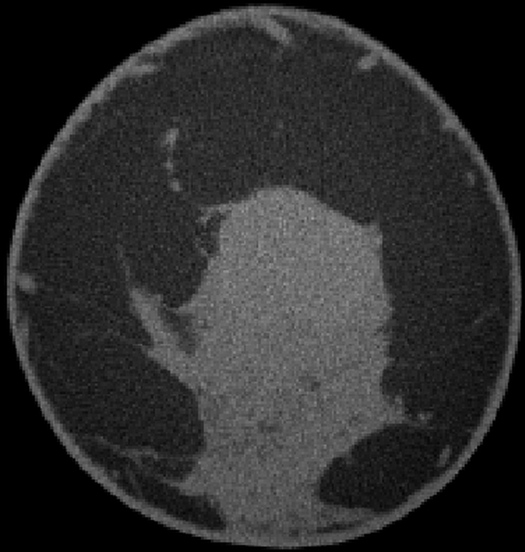
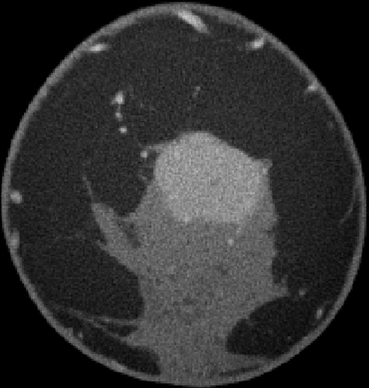
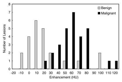
Results: Fifty-four lesions (25 benign, 29 malignant) in 46 subjects were analyzed. Malignant lesions were seen significantly better at contrast-enhanced breast CT than at unenhanced breast CT (P < .001) or mammography (P < .001). Malignant calcifications (malignant lesions manifested mammographically as microcalcifications only, n = 7) were seen better at contrast-enhanced breast CT than at unenhanced breast CT (P < .001) and were seen similarly at contrast-enhanced breast CT and mammography. Malignant lesions enhanced 55.9 HU +/- 4.0 (standard error), whereas benign lesions enhanced 17.6 HU +/- 6.1 (P < .001). Ductal carcinoma in situ (n = 5) enhanced a mean of 59.6 HU +/- 2.8. Receiver operating characteristic curve analysis of lesion enhancement yielded an area under the receiver operating characteristic curve of 0.876.

Conclusion: Conspicuity of malignant breast lesions, including ductal carcinoma in situ, is significantly improved at contrast-enhanced breast CT. Quantifying lesion enhancement may aid in the detection and diagnosis of breast cancer.


Conflict of interest statement

See Materials and Methods for pertinent disclosures.
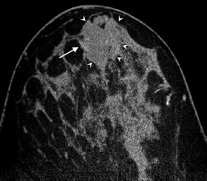
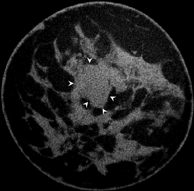
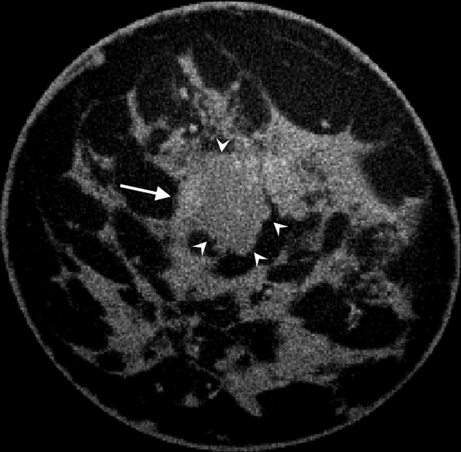
Figures

Figure 1a:
Figure 1a:
Images obtained in a 48-year-old woman. (a) Implant-displaced craniocaudal mammogram shows a mostly obscured mass (arrows). (b) Pre- and (c) postcontrast coronal breast CT images show the same invasive mammary carcinoma mass that enhanced 76.5 HU.
Figure 1b:
Figure 1b:
Images obtained in a 48-year-old woman. (a) Implant-displaced craniocaudal mammogram shows a mostly obscured mass (arrows). (b) Pre- and (c) postcontrast coronal breast CT images show the same invasive mammary carcinoma mass that enhanced 76.5 HU.
Figure 1c:
Figure 1c:
Images obtained in a 48-year-old woman. (a) Implant-displaced craniocaudal mammogram shows a mostly obscured mass (arrows). (b) Pre- and (c) postcontrast coronal breast CT images show the same invasive mammary carcinoma mass that enhanced 76.5 HU.
Figure 2a:
Figure 2a:
Images obtained in a 57-year-old woman. (a) Mediolateral oblique magnification mammogram shows a group of indeterminate microcalcifications (arrow). (b) Precontrast coronal, (c) postcontrast coronal, (d) postcontrast sagittal, and (e) postcontrast transverse breast CT images show the same focus of DCIS (arrow) that enhanced 50.2 HU.
Figure 2b:
Figure 2b:
Images obtained in a 57-year-old woman. (a) Mediolateral oblique magnification mammogram shows a group of indeterminate microcalcifications (arrow). (b) Precontrast coronal, (c) postcontrast coronal, (d) postcontrast sagittal, and (e) postcontrast transverse breast CT images show the same focus of DCIS (arrow) that enhanced 50.2 HU.
Figure 2c:
Figure 2c:
Images obtained in a 57-year-old woman. (a) Mediolateral oblique magnification mammogram shows a group of indeterminate microcalcifications (arrow). (b) Precontrast coronal, (c) postcontrast coronal, (d) postcontrast sagittal, and (e) postcontrast transverse breast CT images show the same focus of DCIS (arrow) that enhanced 50.2 HU.
Figure 2d:
Figure 2d:
Images obtained in a 57-year-old woman. (a) Mediolateral oblique magnification mammogram shows a group of indeterminate microcalcifications (arrow). (b) Precontrast coronal, (c) postcontrast coronal, (d) postcontrast sagittal, and (e) postcontrast transverse breast CT images show the same focus of DCIS (arrow) that enhanced 50.2 HU.
Figure 2e:
Figure 2e:
Images obtained in a 57-year-old woman. (a) Mediolateral oblique magnification mammogram shows a group of indeterminate microcalcifications (arrow). (b) Precontrast coronal, (c) postcontrast coronal, (d) postcontrast sagittal, and (e) postcontrast transverse breast CT images show the same focus of DCIS (arrow) that enhanced 50.2 HU.
Figure 3:
Figure 3:
Bar graph shows enhancement of benign and malignant breast lesions.
Figure 4a:
Figure 4a:
(a) Postcontrast transverse, (b) precontrast coronal, and (c) postcontrast coronal breast CT images of a mucinous adenocarcinoma (arrowheads) with partial rim enhancement (arrow) in a 46-year-old woman.
Figure 4b:
Figure 4b:
(a) Postcontrast transverse, (b) precontrast coronal, and (c) postcontrast coronal breast CT images of a mucinous adenocarcinoma (arrowheads) with partial rim enhancement (arrow) in a 46-year-old woman.
Figure 4c:
Figure 4c:
(a) Postcontrast transverse, (b) precontrast coronal, and (c) postcontrast coronal breast CT images of a mucinous adenocarcinoma (arrowheads) with partial rim enhancement (arrow) in a 46-year-old woman.

Comment in

References

    1. American Cancer Society Breast cancer facts and figures 2009-2010 Atlanta, Ga: American Cancer Society, 2010
    1. Lindfors KK, Boone JM, Nelson TR, Yang K, Kwan AL, Miller DF. Dedicated breast CT: initial clinical experience. Radiology 2008;246(3):725–733 - PMC - PubMed
    1. Newell D, Nie K, Chen JH, et al. Selection of diagnostic features on breast MRI to differentiate between malignant and benign lesions using computer-aided diagnosis: differences in lesions presenting as mass and non-mass-like enhancement. Eur Radiol 2010;20(4):771–781 - PMC - PubMed
    1. Chen SC, Carton AK, Albert M, Conant EF, Schnall MD, Maidment AD. Initial clinical experience with contrast-enhanced digital breast tomosynthesis. Acad Radiol 2007;14(2):229–238 - PMC - PubMed
    1. Perrone A, Lo Mele L, Sassi S, et al. MDCT of the breast. AJR Am J Roentgenol 2008;190(6):1644–1651 - PubMed