. 2010 Aug 18;30(33):11004-10.
doi: 10.1523/JNEUROSCI.1930-10.2010.

The alpha-syntrophin PH and PDZ domains scaffold acetylcholine receptors, utrophin, and neuronal nitric oxide synthase at the neuromuscular junction

Affiliations

The alpha-syntrophin PH and PDZ domains scaffold acetylcholine receptors, utrophin, and neuronal nitric oxide synthase at the neuromuscular junction

Marvin E Adams et al. J Neurosci. .

Abstract

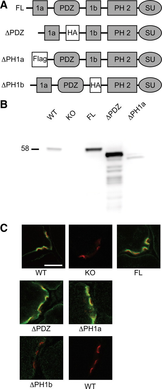
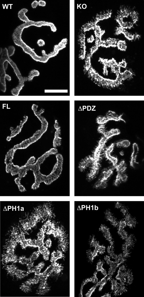
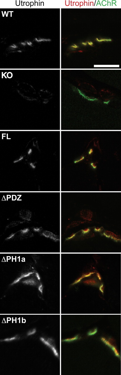
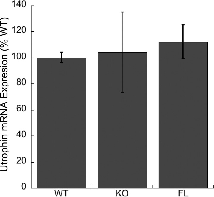
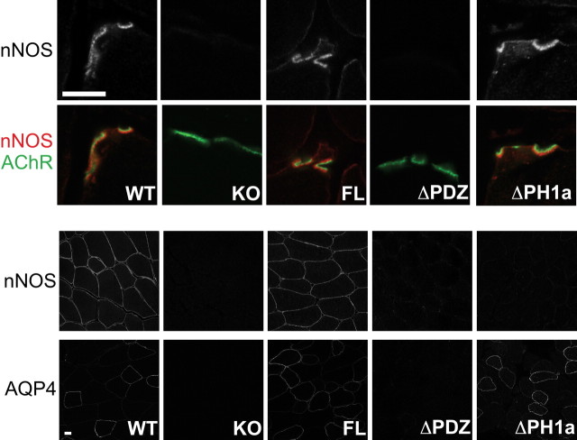
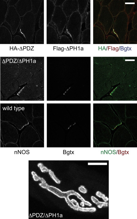
At the neuromuscular junction (NMJ), the dystrophin protein complex provides a scaffold that functions to stabilize acetylcholine receptor (AChR) clusters. Syntrophin, a key component of that scaffold, is a multidomain adapter protein that links a variety of signaling proteins and ion channels to the dystrophin protein complex. Without syntrophin, utrophin and neuronal nitric oxide synthase mu (nNOSmu) fail to localize to the NMJ and the AChRs are distributed abnormally. Here we investigate the contribution of syntrophin domains to AChR distribution and to localization of utrophin and nNOSmu at the NMJ. Transgenic mice expressing alpha-syntrophin lacking portions of the first pleckstrin homology (PH) domain (DeltaPH1a or DeltaPH1b) or the entire PDZ domain (DeltaPDZ) were bred onto the alpha-syntrophin null background. As expected the DeltaPDZ transgene did not restore the NMJ localization of nNOS. The DeltaPH1a transgene did restore postsynaptic nNOS but surprisingly did not restore sarcolemmal nNOS (although sarcolemmal aquaporin-4 was restored). Mice lacking the alpha-syntrophin PDZ domain or either half of the PH1 domain were able to restore utrophin to the NMJ but did not correct the aberrant AChR distribution of the alpha-syntrophin knock-out mice. However, mice expressing both the transgenic DeltaPDZ and the transgenic DeltaPH1a constructs did restore normal AChR distribution, demonstrating that both domains are required but need not be confined within the same protein to function. We conclude that the PH1 and PDZ domains of alpha-syntrophin work in concert to facilitate the localization of AChRs and nNOS at the NMJ.


Figures

Figure 1.
Figure 1.
Generation of syntrophin transgenic mice. A, The protein domains of syntrophin are depicted in the four constructs used to generate transgenic mice. FL is the full-length α-syntrophin transgene. The first pleckstrin homology (PH) domain of syntrophin is naturally split by the PDZ domain into PH1a and PH1b. To generate the ΔPDZ transgene the PDZ domain was replaced by a hemagglutinin epitope tag (HA). In the ΔPH1a and ΔPH1b constructs, the 1a and 1b portions of the first PH domain were replaced by Flag and HA epitope tags, respectively. SU refers to the syntrophin unique region. B, Western blot analysis shows the relative expression levels of α-syntrophin in muscle isolated from the transgenic mice. Note the reduced size of syntrophins missing domains. WT, Wild type; KO, α-syntrophin knock-out. C, Immunofluorescence of NMJs in transverse sections of quadriceps muscle. Fluorescent α-bungarotoxin (red) was used to label the postsynaptic acetylcholine receptors. α-Syntrophin is labeled in green. The ΔPH1b sample (and wild-type control) was labeled using the HA antibody since the α-syntrophin-specific antibody epitope is absent in this construct. Scale bar, 10 μm.
Figure 2.
Figure 2.
Transgenic rescue of aberrant NMJ structure in the α-syntrophin KO muscle. En face views of NMJs from the sternomastoid muscle visualized using fluorescent α-bungarotoxin. The spikes observed in the α-syntrophin KO mice are not present in the FL transgenic, which has continuous smooth edges similar to those of the WT mouse. NMJs of mice lacking the PDZ or either portion of the PH1 domain resemble the KO with spicules and fragmented edges. Scale bar, 20 μm.
Figure 3.
Figure 3.
Postsynaptic utrophin is rescued by all transgenic mice. Immunofluorescence of quadriceps transverse sections shows that postsynaptic utrophin (left panel and red right panel) is present in all transgenic mice whereas it is absent in the α-syntrophin KO. α-Bungarotoxin labeling of acetylcholine receptors (AChR) is shown in green. Scale bar, 10 μm.
Figure 4.
Figure 4.
Utrophin-A mRNA expression in mouse muscle. Real-time PCR of mRNA derived from quadriceps muscle of wild type (WT), α-syntrophin knock-out (KO), and the full-length syntrophin transgenic (FL) shows similar levels of expression in each of these mice. Error bars are SDs with n = 3 mice.
Figure 5.
Figure 5.
The ΔPH1a transgenic mouse rescues nNOS expression at the NMJ but not on the sarcolemma. Immunofluorescence of quadriceps transverse sections shows that nNOS is absent from the NMJs of the KO and ΔPDZ mice. NMJ nNOS expression is rescued by both the full-length and ΔPH1a transgenes. However, sarcolemmal nNOS is rescued only by the full-length transgene, not by the ΔPH1a transgene. Another sarcolemmal protein, aquaporin-4 (AQP4) is rescued by the ΔPH1a transgene. Red, nNOS; AChR (green), α-Bungarotoxin-labeled acetylcholine receptors. Scale bar, 10 μm.
Figure 6.
Figure 6.
NMJ structure is rescued when both the ΔPH1a and ΔPDZ transgenes are present. We generated mice expressing both the ΔPH1a and ΔPDZ transgene as demonstrated by immunofluorescent labeling using antibodies to the epitope tags present in each construct (see Fig. 1). Like the muscles of mice expressing only the ΔPH1a transgene, the ΔPDZ/ΔPH1a muscles express nNOS only at the NMJ and not on the sarcolemma. An en face view of the NMJ (labeled using fluorescent α-bungarotoxin) shows that the NMJs of these mice have smooth continuous edges and lack the radiating spikes seen in the α-syntrophin knock-out mouse and in mice expressing only one of the parent transgenes (see Fig. 2). Scale bar, 20 μm.

Similar articles

Cited by

References

    1. Adams ME, Kramarcy N, Krall SP, Rossi SG, Rotundo RL, Sealock R, Froehner SC. Absence of alpha-syntrophin leads to structurally aberrant neuromuscular synapses deficient in utrophin. J Cell Biol. 2000;150:1385–1398. - PMC - PubMed
    1. Adams ME, Mueller HA, Froehner SC. In vivo requirement of the alpha-syntrophin PDZ domain for the sarcolemmal localization of nNOS and aquaporin-4. J Cell Biol. 2001;155:113–122. - PMC - PubMed
    1. Adams ME, Tesch Y, Percival JM, Albrecht DE, Conhaim JI, Anderson K, Froehner SC. Differential targeting of nNOS and AQP4 to dystrophin-deficient sarcolemma by membrane-directed alpha-dystrobrevin. J Cell Sci. 2008;121:48–54. - PubMed
    1. Ahn AH, Kunkel LM. Syntrophin binds to an alternatively spliced exon of dystrophin. J Cell Biol. 1995;128:363–371. - PMC - PubMed
    1. Alessi A, Bragg AD, Percival JM, Yoo J, Albrecht DE, Froehner SC, Adams ME. gamma-Syntrophin scaffolding is spatially and functionally distinct from that of the alpha/beta syntrophins. Exp Cell Res. 2006;312:3084–3095. - PubMed

Publication types

MeSH terms

LinkOut - more resources