. 2010 Nov;299(5):C1180-94.
doi: 10.1152/ajpcell.00028.2010. Epub 2010 Aug 18.

Ionic currents in intimal cultured synoviocytes from the rabbit

Affiliations

Ionic currents in intimal cultured synoviocytes from the rabbit

R J Large et al. Am J Physiol Cell Physiol. 2010 Nov.

Abstract

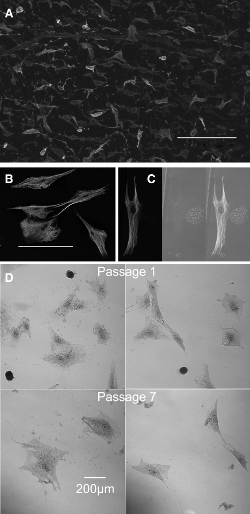
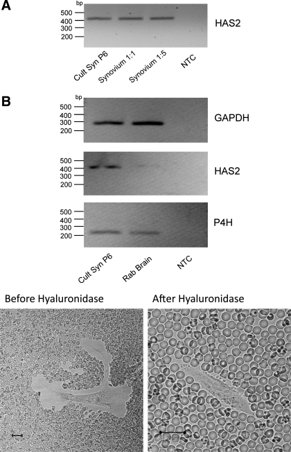
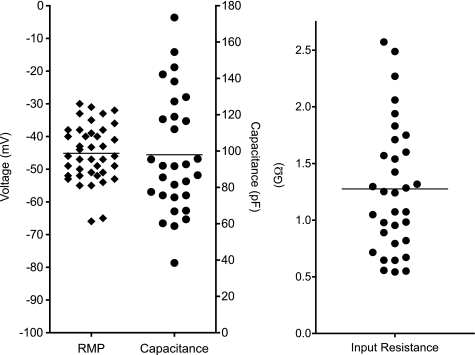
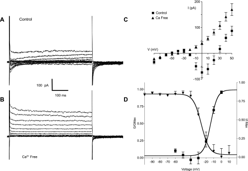
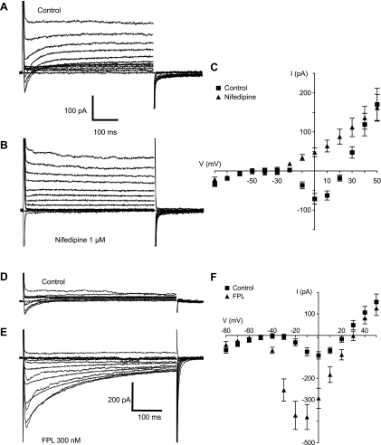
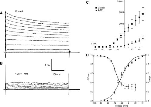
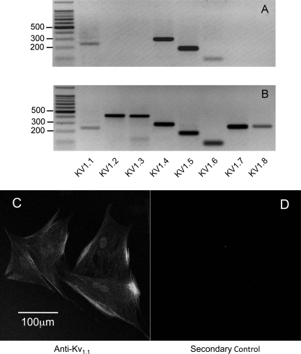
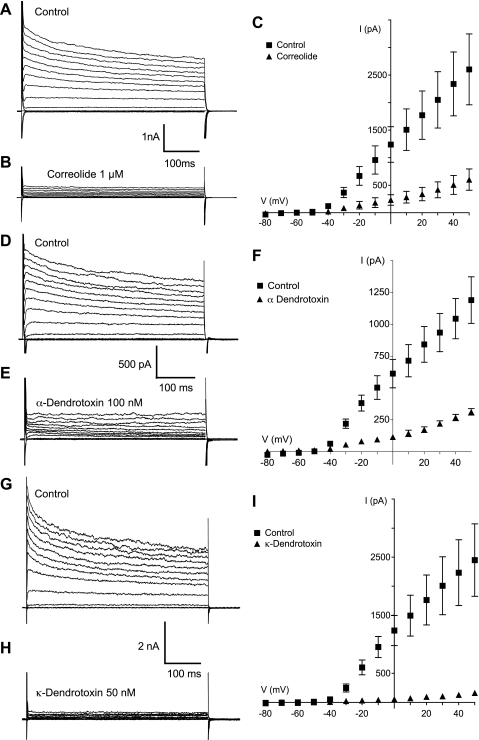
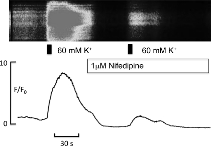
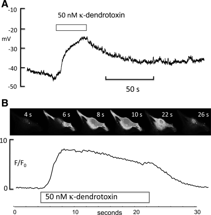
Hyaluronan, a joint lubricant and regulator of synovial fluid content, is secreted by fibroblast-like synoviocytes lining the joint cavity, and secretion is greatly stimulated by Ca(2+)-dependent protein kinase C. This study aimed to define synoviocyte membrane currents and channels that may influence synoviocyte Ca(2+) dynamics. Resting membrane potential ranged from -30 mV to -66 mV (mean -45 ± 8.60 mV, n = 40). Input resistance ranged from 0.54 GΩ to 2.6 GΩ (mean 1.28 ± 0.57 GΩ; ν = 33). Cell capacitance averaged 97.97 ± 5.93 pF. Voltage clamp using C(s+) pipette solution yielded a transient inward current that disappeared in Ca(2+)-free solutions and was blocked by 1 μM nifedipine, indicating an L-type calcium current. The current was increased fourfold by the calcium channel activator FPL 64176 (300 nM). Using K(+) pipette solution, depolarizing steps positive to -40 mV evoked an outward current that showed kinetics and voltage dependence of activation and inactivation typical of the delayed rectifier potassium current. This was blocked by the nonspecific delayed rectifier blocker 4-aminopyridine. The synoviocytes expressed mRNA for four Kv1 subtypes (Kv1.1, Kv1.4, Kv1.5, and Kv1.6). Correolide (1 μM), margatoxin (100 nM), and α-dendrotoxin block these Kv1 subtypes, and all of these drugs significantly reduced synoviocyte outward current. The current was blocked most effectively by 50 nM κ-dendrotoxin, which is specific for channels containing a Kv1.1 subunit, indicating that Kv1.1 is critical, either as a homomultimeric channel or as a component of a heteromultimeric Kv1 channel. When 50 nM κ-dendrotoxin was added to current-clamped synoviocytes, the cells depolarized by >20 mV and this was accompanied by an increase in intracellular calcium concentration. Similarly, depolarization of the cells with high external potassium solution caused an increase in intracellular calcium, and this effect was greatly reduced by 1 μM nifedipine. In conclusion, fibroblast-like synoviocytes cultured from the inner synovium of the rabbit exhibit voltage-dependent inward and outward currents, including Ca(2+) currents. They thus express ion channels regulating membrane Ca(2+) permeability and electrochemical gradient. Since Ca(2+)-dependent kinases are major regulators of synovial hyaluronan secretion, the synoviocyte ion channels are likely to be important in the regulation of hyaluronan secretion.


Figures

Fig. 1.
Fig. 1.
A: whole mount of synovial intima stained with antibody against vimentin. The apparent differences in intensity of staining are due to the relative position in the confocal stack of the vimentin-stained cells. These varied in morphology from ovoid through fibroblastoid to stellate. The horizontal bars in each case indicate 200 μm. B and C: cultured cells from the first passage also stained with antibody against vimentin. B: confocal image of stellate cells rich in vimentin filaments, taken to be type B synoviocytes. C: vimentin-stained cell under laser illumination (left), a smaller rounded cell under bright field illumination (middle), and an overlay of both (right). No evidence of vimentin staining was seen in the small rounded cells, which we took to be macrophages. This conclusion was reinforced by nonspecific esterase (NSE) staining. D: the small rounded cells were strongly NSE positive while the larger cells were negative (top). When the staining was repeated in cells taken from passage 7, no NSE-positive cells were found (bottom).
Fig. 2.
Fig. 2.
Cells from passage 6 (the passage used for electrophysiological studies) were subjected to total RNA extraction using the RNeasy Micro Kit. Total RNA was also prepared from freshly microdissected synovium using the TRIzol method. A: the transcription product was amplified with primers specific for hyaluronan synthase 2 (HAS2), and the resulting DNA bands are shown. HAS2 message was evident in both the passage 6-cultured synoviocytes (Cult Syn P6) and in intact synovium at dilutions of 1:1 and 1:5 but absent from the nontemplate control (NTC). B, bottom: fixed erythrocyte exclusion test. Under normal conditions the synoviocytes were surrounded by a clear area from which erythrocytes were excluded. This clear area disappeared after hyaluronidase addition, suggesting that it was due to hyaluronan secretion by the synoviocyte. Rab, rabbit; P4H, prolyl 4-hydroxylase. Black calibration bar represents 20 μm in each case.
Fig. 3.
Fig. 3.
The range of membrane potential measured in zero current clamp mode is shown for 40 cells. Values ranged from −30 mV to −66 mV with a mean of −45 mV. There was no clear bimodal distribution, indicating a fairly homogenous cell population. The measured capacitance ranged from <40 to ∼180 pF, with a mean of 97.97 pF, reflecting the large variations in surface area of cultured cells. The mean input resistance was 1.27 GΩ with a range of 0.54 to 2.6 GΩ. RMP, resting membrane potential.
Fig. 4.
Fig. 4.
A: family of currents elicited by holding the cell at −80 mV and stepping through a series of voltages from −80 mV to +50 mV in normal Hanks' solution. K+ ions in the pipette solution were replaced with Cs+ to block K+ currents. At voltages positive to −20 mV, a transient inward current was evoked that peaked within 5 ms and relaxed to 0 within 200 ms. This current was partially offset by an outward current that activated over the same voltage range and was likely to be due to cesium ions being carried through potassium channels. B: same protocol as in A applied to the same cell but this time in Ca2+ -free Hanks' solution. The transient inward current was abolished, leaving only the outward current carried by Cs+. C: current-voltage (I-V) relationship of the inward current is summarized in the plot of the mean results of six such experiments. Peak inward current (■) developed at voltages positive to −20 mV, reached a maximum at 0 mV, and reversed at just under +30 mV. In Ca2+-free Hanks' solution (▲) the inward current was abolished at all voltages. D: voltage dependence of activation (■) and inactivation (▲) of the inward current. The mean results of 14 experiments were fitted with a Boltzmann equation giving a half-maximal voltage (V1/2) of activation of −14 ± 1.7 mV. The current activated at voltages positive to −10 mV and was maximally active at voltages positive to 0 mV. Voltage-dependent inactivation was investigated by holding cells at a series of conditioning potentials from −100 mV to +10 mV for 2 s before stepping to a test potential of 0 mV for 500 ms. For analysis, the peak current at 0 mV was measured at each conditioning potential, normalized to the maximum current (Imax), and plotted against the appropriate potential. Mean data points in 5 such experiments were again fitted with a Boltzmann equation, yielding a V1/2 of inactivation of −26 ± 1.68 mV. It is clear from this plot that availability of the inward current was high over the measured range of resting membrane potential. Gmax, maximum conductance.
Fig. 5.
Fig. 5.
A and B: family of currents (evoked by the same protocol as that described in Fig. 4) before (A) and in the presence of nifedipine (B). The transient inward current was blocked, indicating that it was an L-type calcium current. C: summary of 6 such experiments. Mean inward current (■) peaked at 0 mV and reversed at +23 mV. In the presence of nifedipine, inward current was abolished, leaving only the outward current carried by Cs+. D and E: effects of the calcium channel agonist FPL-64176. The control inward currents (D) were greatly enhanced by 300 nM FPL 64176 (FPL; E). This was accompanied by somewhat slower activation kinetics and by very much slower inactivation, such that the currents were not fully inactivated at the end of the 500-ms sweep. F: a summary of 12 such experiments showing that the mean peak inward current increased in amplitude and that the voltage at which current peaked shifted negatively by 10 mV. Peak inward current increased by 308% from a control value (■) of 93 ± 17 pA to 380 ± 58 pA in the presence of FPL (▲; n = 12; P < 0.001).
Fig. 6.
Fig. 6.
A: family of outward currents evoked by the protocol described previously but using K+ pipettes. The outward current activated rapidly and then relaxed slowly throughout the remainder of the 500-ms test pulse. B: the currents were greatly attenuated in the presence of 1 mM 4-aminopyridine (4-AP). C: summary of the current-voltage plot for eight experiments in control conditions (■) and in the presence of 4-AP (▲). The current activated at −50 mV and showed outward rectification upon stepping to more positive potentials. 4-AP reduced the maximum current by 73% (n = 8, P < 0.0001). D: current activation and inactivation properties. The current activated (■) at potentials positive to −80 mV and was maximally active at +40 mV. V1/2 of activation was −21 ± 0.7 mV (n = 38). An inactivation protocol (conditioning pulses of 2-s duration in the range −100 to +20 mV stepping to a test potential of +40 mV) yielded the inactivation curve (▲). Inactivation began at potentials positive to −70 mV and reached a plateau at about 0 mV although 30% of the current was still available.
Fig. 7.
Fig. 7.
Kv1 subunit expression in cDNA from passage 6 synoviocytes. A: products of appropriate size were obtained for Kv1.1, Kv1.4, Kv1.5, and Kv1.6 following 35 cycles of PCR amplification (n = 3). B: as a positive control, cDNA from rabbit brain was examined using the same primers. All eight Kv1 subtypes were found in rabbit brain. C and D: immunostaining with a mouse monoclonal antibody against Kv1.1 (C) and the control experiment with only secondary antibody present (D).
Fig. 8.
Fig. 8.
Outward currents under control conditions are shown in A, D, and G while those in the presence of correolide (1 μM), α-dendrotoxin (100 nM), and κ-dendrotoxin (50 nM) are shown in B, E, and H. respectively. The most potent blocking effect by far was that of κ-dendrotoxin, which almost completely eliminated the current. The results for each set of experiments are summarized in the current-voltage plots of C, F, and I.
Fig. 9.
Fig. 9.
Top: pseudo linescan of a synoviocyte loaded with the calcium indicator fluo-4 (see materials and methods); shown are the effects of increasing external K+ concentration for 5 s (indicated by the black bar) before and in the presence of 1 μM nifedipine. Bottom: F/F0 plot of the same experiment. Depolarization of the membrane with high potassium caused a large increase in intracellular calcium, and this effect was greatly attenuated in the presence of nifedipine.
Fig. 10.
Fig. 10.
A: synoviocyte in current clamp with a resting membrane potential between −50 and −40 mV. When 50 nM κ-dendrotoxin was added, the cell depolarized by >20 mV, suggesting a role for Kv1.1 channels in the maintenance of resting membrane potential. B: montage of a fluo-4-loaded synoviocyte to which 50 nM κ-dendrotoxin was added after 4 s. Intracellular calcium levels rose to a peak between 8 and 10 s, and this was well maintained until the toxin was removed, whereupon it fell to control levels as shown by the F/F0 plot below.

Similar articles

Cited by

References

    1. Anggiansah CL, Scott D, Poli A, Coleman PJ, Badrick E, Mason RM, Levick JR. Regulation of hyaluronan secretion into rabbit synovial joints in vivo by protein kinase C. J Physiol 550: 631–640, 2003 - PMC - PubMed
    1. Aune TM. Inhibition of arthritis by L-type calcium channel antagonists nimodipine, nisoldipine and nifedipine. US patent 5478848, 1995
    1. Badou A, Jha MK, Matza D, Mehal WZ, Freichel M, Flockerzi V, Flavell RA. Critical role for the beta regulatory subunits of Cav channels in T lymphocyte function. Proc Natl Acad Sci USA 103: 15529–15534, 2006 - PMC - PubMed
    1. Bae YM, Kim A, Kim J, Park SW, Kim TK, Lee YR, Kim B, Cho SI. Serotonin depolarizes the membrane potential in rat mesenteric artery myocytes by decreasing voltage-gated K+ currents. Biochem Biophys Res Commun 347: 468–476, 2006 - PubMed
    1. Baumgarten L, Villereal M. Bradykinin stimulates Ca2+ entry via nitrendipine-sensitive Ca2+ channels in cultured human fibroblasts. Agents Actions Suppl 38: 1–8, 1992 - PubMed

Publication types

MeSH terms