. 2010 Sep 15;185(6):3369-78.
doi: 10.4049/jimmunol.0902541. Epub 2010 Aug 18.

Proximal events in 7,12-dimethylbenz[a]anthracene-induced, stromal cell-dependent bone marrow B cell apoptosis: stromal cell-B cell communication and apoptosis signaling

Affiliations

Proximal events in 7,12-dimethylbenz[a]anthracene-induced, stromal cell-dependent bone marrow B cell apoptosis: stromal cell-B cell communication and apoptosis signaling

Jessica E Teague et al. J Immunol. .

Abstract

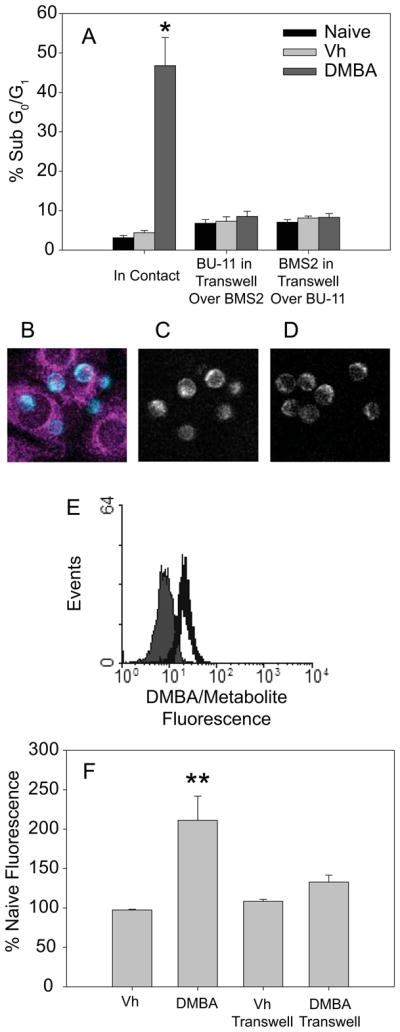
Intercellular communication is an essential process in stimulating lymphocyte development and in activating and shaping an immune response. B cell development requires cell-to-cell contact with and cytokine production by bone marrow stromal cells. However, this intimate relationship also may be responsible for the transfer of death-inducing molecules to the B cells. 7,12-Dimethylbenz[a]anthracene (DMBA), a prototypical polycyclic aromatic hydrocarbon, activates caspase-3 in pro/pre-B cells in a bone marrow stromal cell-dependent manner, resulting in apoptosis. These studies were designed to examine the hypothesis that an intrinsic apoptotic pathway is activated by DMBA and that the ultimate death signal is a DMBA metabolite generated by the stromal cells and transferred to the B cells. Although a loss of mitochondrial membrane potential did not occur in the DMBA/stromal cell-induced pathway, cytochrome c release was stimulated in B cells. Caspase-9 was activated, and formation of the apoptosome was required to support apoptosis, as demonstrated by the suppression of death in Apaf-1(fog) mutant pro-B cells. Investigation of signaling upstream of the mitochondria demonstrated an essential role for p53. Furthermore, DMBA-3,4-dihydrodiol-1,2-epoxide, a DNA-reactive metabolite of DMBA, was sufficient to upregulate p53, induce caspase-9 cleavage, and initiate B cell apoptosis in the absence of stromal cells, suggesting that production of this metabolite by the stromal cells and transfer to the B cells are proximal events in triggering apoptosis. Indeed, we provide evidence that metabolite transfer from bone marrow stromal cells occurs through membrane exchange, which may represent a novel communication mechanism between developing B cells and stromal cells.


Figures

Fig. 1
Fig. 1
DMBA induces caspase-independent mitochondrial cytochrome c release, but not membrane potential loss, in B cells co-cultured with bone marrow stromal cells. (A) BU-11/BMS2 co-cultures were treated with vehicle (Vh, 0.1% DMSO) or DMBA (1 μM), and BU-11 cells were harvested after 2–8 hr. Cytochrome c release was analyzed by immunoblotting of cytoplasmic extracts from digitonin-permeabilized cells. Data are representative of 3 experiments. (B) BU-11/BMS2 co-cultures were treated with Vh or DMBA (1 μM) or BU-11 cultures were treated with Vh or etoposide (0.1 μg/ml) (inset) for 4–10 hrs. BU-11 cells were analyzed for mitochondrial membrane potential loss by JC-1 staining followed by flow cytometry. Data are are presented as means + SE from at least three experiments. (C) BU-11/BMS2 co-cultures were pre-treated with Vh or the pan-caspase inhibitor VAD-FMK (30 μM) for 30 min prior to treatment with Vh or DMBA (1 μM) for 8 hr. Cytochrome c release was analyzed as above. Data are representative of 3 experiments. *Statistically greater than Vh treated (p<0.05, ANOVA, Tukey-Kramer).
Fig. 2
Fig. 2
The apoptosome is essential for DMBA/stromal cell-induced B cell apoptosis. BU-11/BMS2 co-cultures were treated with Vh or DMBA (1 μM), and BU-11 cells were harvested after 2–10 hr. (A) Total proteins were extracted and analyzed for active caspase-9 fragments and for β-actin by immunoblotting. Data are representative of 4 experiments. (B) Cytosolic proteins were extracted, and caspase-9-like activity was measured using p-nitroaniline-conjugated LEHD substrate. Data were quantified as the average fold increase in caspase-9-like activity relative to the activity in untreated cells and presented as means ±SE from 4 experiments. (C) Primary proB cells isolatedfrom mice either homozygous or heterozygous for the Apaf-1fogmutation, were treated in co-culture with BMS2 with Vh or DMBA (1 μM). Pro-B cells were analyzed for apoptosis by PI staining 16 hr after treatment. The percentage of death measured in naïve cell populationswas subtracted prior to analysis. Data are presented as means + SE from primary pro-B cells prepared from 5–6 individual mice. *Statistically different from Vh (p<0.05, ANOVA, Dunnett’s).* *Statistically greater than all other groups(p<0.05, ANOVA, T ukey-Kramer).
Fig. 3
Fig. 3
Contribution of p53 to DMBA/stromal cell-induced B cell apoptosis. (A) BU-11/BMS2 co-cultures were treated with Vh or DMBA (1 μM), and BU-11 cells were harvested after 2–10 hr. Total proteins were extracted and analyzed for p53 and for β-actin by immunoblotting. Data are representative of 3 experiments. (B, C) Primary pro-B cells isolated from wildtype or p53 mutant mice were treated in co-culture with BMS2 cells with Vh or DMBA (1 μM) for 2–16 hr. (B) Pro-B cells were analyzed for apoptosis by PI staining 16 hr after treatment. The percentage of death measured in naïve cells was subtracted prior to analysis. Data are presented as means + SE from primary pro-B cells prepared from 3–4 individual mice. (C) Total proteins were extracted from pro-B cells and analyzed for cleaved caspase-3 and for β-actin by immunoblotting. A caspase-3 positive control was included on each gel to enable comparison between blots. Data are representative of experiments using primary pro-B cells from 3 individual mice. *Statistically greater than all other treatment groups (p<0.05, ANOVA, Tukey-Kramer). **Statistically less than wildtype DMBA treated (p<0.05, ANOVA, Tukey -Kramer).
Fig. 4
Fig. 4
A terminal metabolite of DMBA induces stromal cell-independent B cell apoptosis. BU-11 cell suspension cultures were treated with Vh or DMBA-DE (1 μM) for the indicated times. (A) BU-11 cells were analyzed for apoptosis by PI staining. The percentage of death measured in naïve cells was subtracted prior to analysis. Data are presented as means + SE from at least 3 experiments. (B) Total proteins were extracted from BU-11 cells and analyzed for p53 and β-actin by immunoblotting. Data are representative of at least 3 experiments. (C) Primary pro-B cells isolated from wildtype orp53 mutant mice were treated with Vh or DMBA-DE (0.01 μM). Pro-B cells were analyzed for apoptosis by PI staining 16 hr after treatment. The percentage of death measured in naïve cells was subtracted prior to analysis. Data are presented as means + SE from primary pro-B cells prepared from 4 individual mice. *Statistically different from Vh (p<0.05, ANOVA, Dunnett’s).
Fig. 5
Fig. 5
Cell contact is required for DMBA/stromal cell-induced apoptosis, potentially facilitating metabolite transfer from BMS2 to BU-11 cells. (A) BMS2 cells were cultured in plates as usual or in 3 μm pore size transwells. BU-11 cells were added directly to BMS2 monolayers or to transwells suspended over BMS2 monolayers. Co-cultures were treated with Vh or DMBA (1 μM) for 16 hr. BU-11 cells were analyzed for apoptosis by PI staining. Data are presented as means + SE from 3 experiments. (B-D) BMS2 cultures were treated with 10 μM DMBA for 30 min, washed, incubated for 1 hr, then washed again. BU-11 cells then were centrifuged onto the BMS2 cell layer and cultures immediately analyzed by two-photon microscopy for DMBA/metabolite fluorescence transfer. (B) False color image of BU-11 cells in contact with the BMS2 cell monolayer overlayed on the BMS2 cell image. (C) Image of BU-11 cells in contact with the BMS2 cell monolayer. (D) BU-11 cells not in contact with the BMS2 cell monolayer. Representative data from one of three similar experiments are presented. BU-11 cells in contact with BMS2 cells tended to contain a higher level of fluorescence than those no it contact with BMS2 (p<0.06). (E, F) BMS2 cultures were treated with Vh or DMBA (10 μM) for 30 min, washed extensively, incubated for 5 hr, and washed again. BU-11 cells then were added directly to the BMS2 cell layer or to a 1 μm pore size transwell insert above the BMS2 cells. BU-11 cells were analyzed at 18 hr by flow cytometry for fluorescence at 450 nm. (E) Representative fluorescence histogram of BU-11 cells cultured on naïve (filled) or DMBA-treated (open) BMS2 cells. (F) DMBA/metabolite fluorescence expressed as percent of naïve fluorescence. Data are presented as the means + SE from 3 experiments. *Statistically greater than other treatment groups (p<0.05, ANOVA, Tukey-Kramer). **Statistically different from naïve and from DMBA in transwell groups (p < 0.05, ANOVA, Tukey-Kramer).
Fig. 6
Fig. 6
B lineage cells take up membrane lipid from bone marrow stromal cells in a contact-dependent manner. BMS2 cells were left unstained or were labeled with Vybrant DiO plasma membrane dye according to the manufacturer’s protocol. (A) BU-11 cells were added directly to unstained or stained adherent BMS2 cells (Contact) or in a 3 μm pore size transwell insert over the BMS2 cells (Transwell) and harvested after 16 hr. In addition, naïve BU-11 cells were resuspended for 20 min in 16 hr supernatants from unstained or DiO-stained BMS2 cells (Supernatant). DiO uptake was analyzed by flow cytometry in live-gated cells. Inset: The filled gray histogram representsdata from BU -11 cells cultured in contact with unstained BMS2 cells, the open gray lined histogram represents data from BU-11 cells cultured in transwells over DiO-stained BMS2 cells, and the open black lined histogram represents data from BU-11 cells cultured in contact with DiO-stained BMS2 cells. (B) BU-11 cells were added directly to unstained or stained adherent BMS2 cells, harvested after 0.5–2 hrs, and analyzed by flow cytometry. (C, D) Primary pro-B cells, splenic B cells, or splenic T cells from wildtype C57Bl/6 mice were added to unstained or DiO-stained BMS2 cells directly, and lymphocytes were assayed 16 hr later by flow cytometry. (C) Representative histograms of lymphocytes on unstained (filled) or DiO-stained (open) BMS2 cells. (D) Quantification of DiO positive lymphocytes. Data are presented as means + SE from 3–4 experiments or from 3–5 experiments with cells from individual mice. *Statistically different from lymphocytes on unstained BMS2 cell controls (p< 0.05, ANOVA, Tukey-Kramer).

References

    1. White KL, Jr, Lysy HH, Holsapple MP. Immunosuppression by polycyclic aromatic hydrocarbons: A structure-activity relationship in B6C3F1 and DBA/2 mice. Immunopharmacology. 1985;9:155–164. - PubMed
    1. Dean JH, Luster MI, Boorman GA, Lauer LD, Luebke RW, Lawson LD. Immunesuppression following exposure of mice to the carcinogen benzo(a)pyrene but not by the noncarcinogenic congener benxo(e)pyrene. Clin Exp Immunol. 1983;52:199. - PMC - PubMed
    1. Dean J, Ward E, Murray M, Lauer L, House R, Stillman W, Hamilton T, Adams D. Immunosuppression following 7,12-deimthylbenz[a]anthracene exposure in B6C3F1--II. Altered cell-mediated immunity and tumore resistance. Int J Immunopharm. 1986;8:189–198. - PubMed
    1. Thurmond L, Lauer L, House R, Cook J, Dean J. Immunosuppression following exposure to 7,12-dimethylbenz[a]anthracene (DMBA) in Ah-responsive and Ah-nonresponsive mice. Toxicology and Applied Pharmacology. 1987;91:450–460. - PubMed
    1. Ward EC, Murray MJ, Lauer LD, House RV, Irons R, Dean JH. Immunosuppression following 7,12-dimethylbenz[a]anthracene exposure in B6C3F1 mice. I. Effect on humoral immunity and host resistance. Toxicol Appl Pharmacol. 1984;75:299–308. - PubMed

Publication types

MeSH terms

Substances