. 2010 Sep;69(9):945-58.
doi: 10.1097/NEN.0b013e3181efa658.

Axonal pathology precedes demyelination in a mouse model of X-linked demyelinating/type I Charcot-Marie Tooth neuropathy

Affiliations

Axonal pathology precedes demyelination in a mouse model of X-linked demyelinating/type I Charcot-Marie Tooth neuropathy

Natalie Vavlitou et al. J Neuropathol Exp Neurol. 2010 Sep.

Abstract

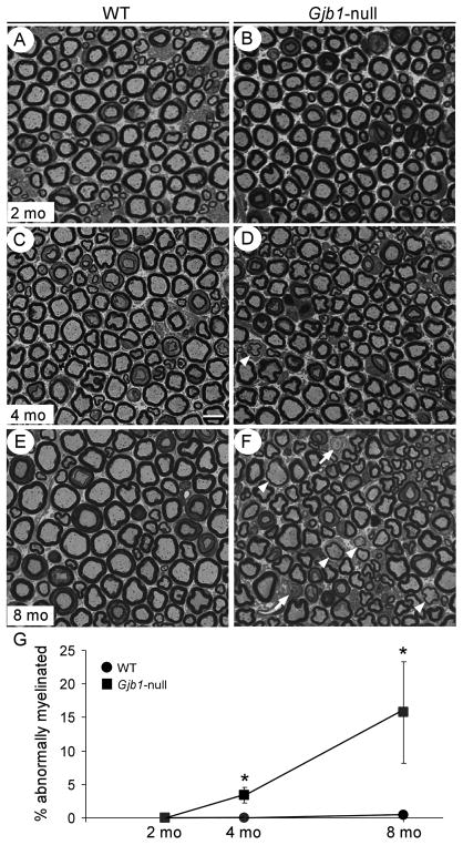
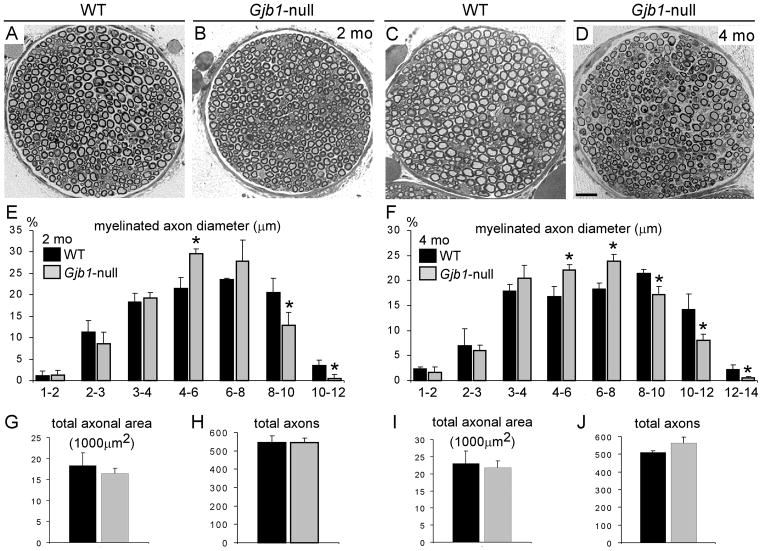
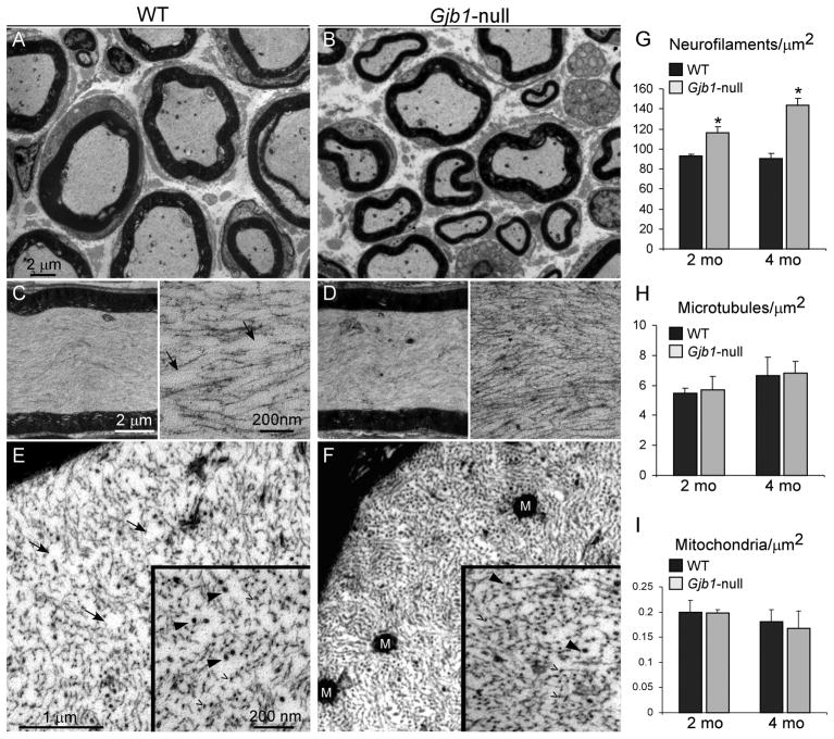
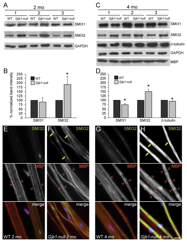
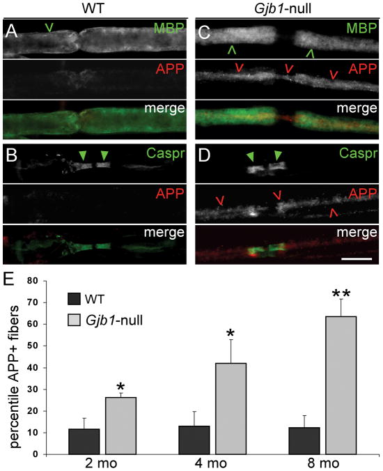
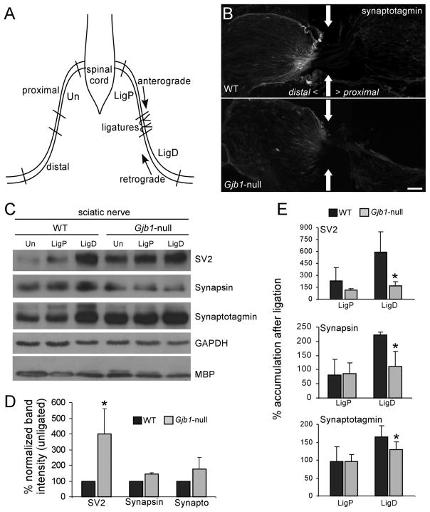
The X-linked demyelinating/type I Charcot-Marie-Tooth neuropathy (CMT1X) is an inherited peripheral neuropathy caused by mutations in GJB1, the gene that encodes the gap junction protein connexin32. Connexin32 is expressed by myelinating Schwann cells and forms gap junctions in noncompact myelin areas, but axonal involvement is more prominent in X-linked compared with other forms of demyelinating Charcot-Marie-Tooth disease. To clarify the cellular and molecular mechanisms of axonal pathology in CMT1X, we studied Gjb1-null mice at early stages (i.e. 2-4 months old) of the neuropathy, when there is minimal or no demyelination. The diameters of large myelinated axons were progressively reduced in Gjb1-null mice compared with those in wild-type littermates. Furthermore, neurofilaments were relatively more dephosphorylated and more densely packed starting at 2 months of age. Increased expression of β-amyloid precursor protein, a marker of axonal damage, was also detected in Gjb1-null nerves. Finally, fast axonal transport, assayed by sciatic nerve ligation experiments, was slower in distal axons of Gjb1-null versus wild-type animals with reduced accumulation of synaptic vesicle-associated proteins. These findings demonstrate that axonal abnormalities including impaired cytoskeletal organization and defects in axonal transport precede demyelination in this mouse model of CMT1X.


Figures

Figure 1
Figure 1
Sciatic nerve demyelination in Gjb1-null mice is minimal up to 4 months of age. (A–F) Semithin sections from 2- (A, B), 4- (C, D) and 8-month-old (E, F) wild type (WT) (A, C, E) and Gjb1-null (B, D, F) mice. In Gjb1-null sciatic nerves there are no demyelinated or remyelinated axons at 2 months of age. Abnormally myelinated axons are rare at 4 months (D), and become more numerous at 8 months (F), including demyelinated (arrows) and remyelinated (arrowheads). Bar = 10 μm. (G) Quantitative analysis of sciatic nerve myelination in WT and Gjb1-null mice at 2, 4, and 8 months of age (n = 4 per genotype and age group) shows very mild demyelination in Gjb1-null mice at 4 months with 3.4 ± 1.2% (average ± SD) of fibers being abnormally myelinated vs. none in WT, (p = 0.01, Mann-Whitney U test); by 8 months there is progression (15.7 ± 7.6% of fibers abnormally myelinated vs. 0.4 ± 0.5% in WT; p = 0.03).
Figure 2
Figure 2
Reduced axonal diameters in Gjb1-null mice. (A–D) Representative semithin sections of femoral motor nerves from 2- (A, B) and 4-month-old (C, D) wild type (WT) (A, C) and Gjb1-null (B, D) mice. A few axons are demyelinated or remyelinated at 4 months of age in Gjb1-null nerves. Scale bar = 20 μm. (E–J) Quantitative analysis of axon diameters (E, F), total axonal area (G, I), and total numbers of axons (H, J) in WT and Gjb1-null mice (n = 3 per age group and genotype) at 2 months (E, G, H) and 4 months (F, I, J). At both ages the percentages of large myelinated fibers (>8μm) is reduced in Gjb1-null vs. WT nerves. In contrast, Gjb1-null nerves show higher percentages of medium sized fibers (4 to 6 μm at 2 months; 4 to 8 μm at 4 months). The total axonal area and the total number of axons are not significantly changed in Gjb1-null nerves, suggesting a shift to smaller diameters without axonal loss. *Indicates significant differences (2-tailed t-test).
Figure 3
Figure 3
Increased density of axonal neurofilaments in Gjb1-null femoral nerves. (A–F) Electron micrographs from femoral motor nerves of 4-month-old wild type (WT) (A, C, E) and Gjb1-null (B, D, F) mice. Low-magnification images (A, B) show that axons are normally myelinated in Gjb1-null nerve. Higher magnification images of longitudinal (C, D) and transverse (E, F) sections of normally myelinated axons illustrate the higher density of neurofilaments (NFs) in the Gjb1-null vs. the WT axons. At higher magnification of longitudinal (insets, C, D) and transverse sections (insets, E, F), NFs (open arrowheads) appear more tightly packed in Gjb1-null axons; microtubules (arrowheads) do not show clear differences in density. Areas devoid of NFs seen in the WT (arrows in C and E) are absent in the Gjb1-null (D, F). M = mitochondria. (G–I) Average density per μm2 of NFs (G), microtubules (H) and mitochondria (I) obtained in at least 10 normally myelinated axons (6-to 8-μm diameter) from 2- and 4-month-old mice in each genotype (n = 3–5 mice per genotype and age group). The density of NFs is higher in Gjb1-null mice vs. WT starting at 2 months and is further increased at 4 months. The densities of microtubules and mitochondria are not significantly different at any age (*Indicates significant results).
Figure 4
Figure 4
Early dephosphorylation of axonal neurofilaments in Gjb1-null nerves without demyelination. (A–D) Immunoblot analysis of cytoskeletal components in sciatic nerves from 3 pairs of 2-month-old (A, B) and 4 month-old (C, D) wild (WT) and Gjb1-null littermates. (A, C) Representative blots are shown for antibodies to phosphorylated (SMI31) and non-phosphorylated (SMI32) NF-heavy (NF-H; 200 kDa) and β-tubulin (55 kDa) and glyceraldehyde 3-phosphate dehydrogenase (GAPDH) (37 kDa, loading control) and myelin basic protein (MBP, 20 kDa). In all pairs, SMI32 levels are greater in Gjb1-null vs. WT littermates. At 2 months SMI31 levels are not different, but at 4 months, SMI31 levels appear reduced, along with minimal reduction of β-tubulin; MBP levels are unchanged. (B, D) Results of quantitative analysis (average band intensity values for cytoskeletal proteins from at least 3 pairs of WT and Gjb1-null mice each run at least 3 times in SDS-PAGE and electrotransferred onto nitrocellulose for immunolabeling, normalized for loading to GAPDH). Non-phosphorylated NF-H levels are significantly greater in Gjb1-null nerves while phosphorylated NF-H levels are decreased at 4 months. β-tubulin levels are mildly reduced in Gjb1-null nerves at 4 months. *Indicates significant results (p < 0.05, paired t-test). (E–H) Immunostaining of sciatic nerve teased fibers for SMI32 (green) and MBP (red); cell nuclei are stained with DAPI (blue). Axons from 2-month-old Gjb1-null mice (F) have more SMI32-immunoreactivity (IR) (green arrows) than those from WT littermates (E), with inhomogeneous pattern along the entire axons. At 4 months, SMI32-IR completely fills some axons (H) of Gjb1-null mice. The SMI32+ axons have normal-appearing, MBP+ myelin sheaths (red open arrowheads). Scale bar in H: 10 μm.
Figure 5
Figure 5
Amyloid precursor protein (APP)+ axons in Gjb1-null sciatic nerves. (A–D) Representative images of teased fibers from 4-month-old wild type (WT) (A, B) and Gjb1-null (C, D) mice, double labeled with antibodies to APP (red) and either myelin basic protein (MBP, green, A, C) or contactin-associated protein (Caspr, green, B, D). Many Gjb1-null axons (red open arrowheads), but not WT axons, are APP+; these are surrounded by normal-appearing MBP+ myelin sheaths (green open arrowheads) and have normal-appearing Caspr+ paranodes (green arrowheads). Scale bar: 10 μm. (E) Quantitative analysis of APP+ axons in 2-, 4-, and 8-month old mice (n = 3 or 4 in each genotype and age group). The percentile of APP+ axons increases with age and, at every age, is significantly higher in Gjb1-null vs. WT mice.
Figure 6
Figure 6
Early impairment of axonal transport in Gjb1-null mice. (A) Schematic of the sciatic nerve ligation experiment. One sciatic nerve was doubly ligated; 3 hours later, segments proximal (LigP) and distal (LigD) to the ligation and the corresponding segments of the contralateral/unligated nerve (Un) were collected. (B) Immunostaining at the distal ligation site (arrows) shows more synaptotagmin accumulation in the wild type (WT) than in the Gjb1-null nerve. Bar = 40 μm. (C) Representative immunoblots of lysates from ligated (LigP and LigD) and unligated nerves, from WT and Gjb1-null mice. Membranes were blotted with monoclonal antibodies to SV2 (95 kDa), synapsin (70 kDa) and synaptotagmin (65 kDa), glyceraldehyde 3-phosphate dehydrogenase (GAPDH, loading control) and myelin basic protein (MBP) (20 kDa). The levels of SV2, synapsin and synaptotagmin in LigD (vs. same region of unligated nerves) are higher in WT than in Gjb1-null nerves; their steady state levels in unligated nerves are higher in the Gjb1-null. GADPH and MBP levels are similar in WT and Gjb1-null mice. (D, E) Quantitative results from 3 independent experiments. (D) Bars represent average percentage increase in synaptic protein levels (normalized for GAPDH signals) in unligated nerves from Gjb1-null vs. WT mice. (E) Bars represent average percentage increase in the synaptic protein levels in LigD and LigP vs. the contralateral unligated nerves, after normalization for GAPDH signals. SV2, synapsin, and synaptotagmin levels are significantly higher in WT vs. the Gjb1-null LigD segments (1-tailed paired t-test).

Similar articles

Cited by

References

    1. Bergoffen J, Scherer SS, Wang S, et al. Connexin mutations in X-linked Charcot-Marie-Tooth disease. Science. 1993;262:2039–42. - PubMed
    1. Scherer SS, Deschênes SM, Xu Y-T, et al. Connexin32 is a myelin-related protein in the PNS and CNS. J Neurosci. 1995;15:8281–94. - PMC - PubMed
    1. Balice-Gordon RJ, Bone LJ, Scherer SS. Functional gap junctions in the Schwann cell myelin sheath. J Cell Biol. 1998;142:1095–1104. - PMC - PubMed
    1. Meier C, Dermietzel R, Davidson KG, et al. Connexin32-containing gap junctions in Schwann cells at the internodal zone of partial myelin compaction and in Schmidt-Lanterman incisures. J Neurosci. 2004;24:3186–98. - PMC - PubMed
    1. Kleopa KA, Scherer SS. Molecular genetics of X-linked Charcot-Marie-Tooth disease. Neuromolecular Med. 2006;8:107–122. - PubMed

Publication types

MeSH terms