. 2010 Dec;62(12):3686-94.
doi: 10.1002/art.27709.

Intraarticular injection of heparin-binding insulin-like growth factor 1 sustains delivery of insulin-like growth factor 1 to cartilage through binding to chondroitin sulfate

Affiliations

Intraarticular injection of heparin-binding insulin-like growth factor 1 sustains delivery of insulin-like growth factor 1 to cartilage through binding to chondroitin sulfate

Rachel E Miller et al. Arthritis Rheum. 2010 Dec.

Abstract

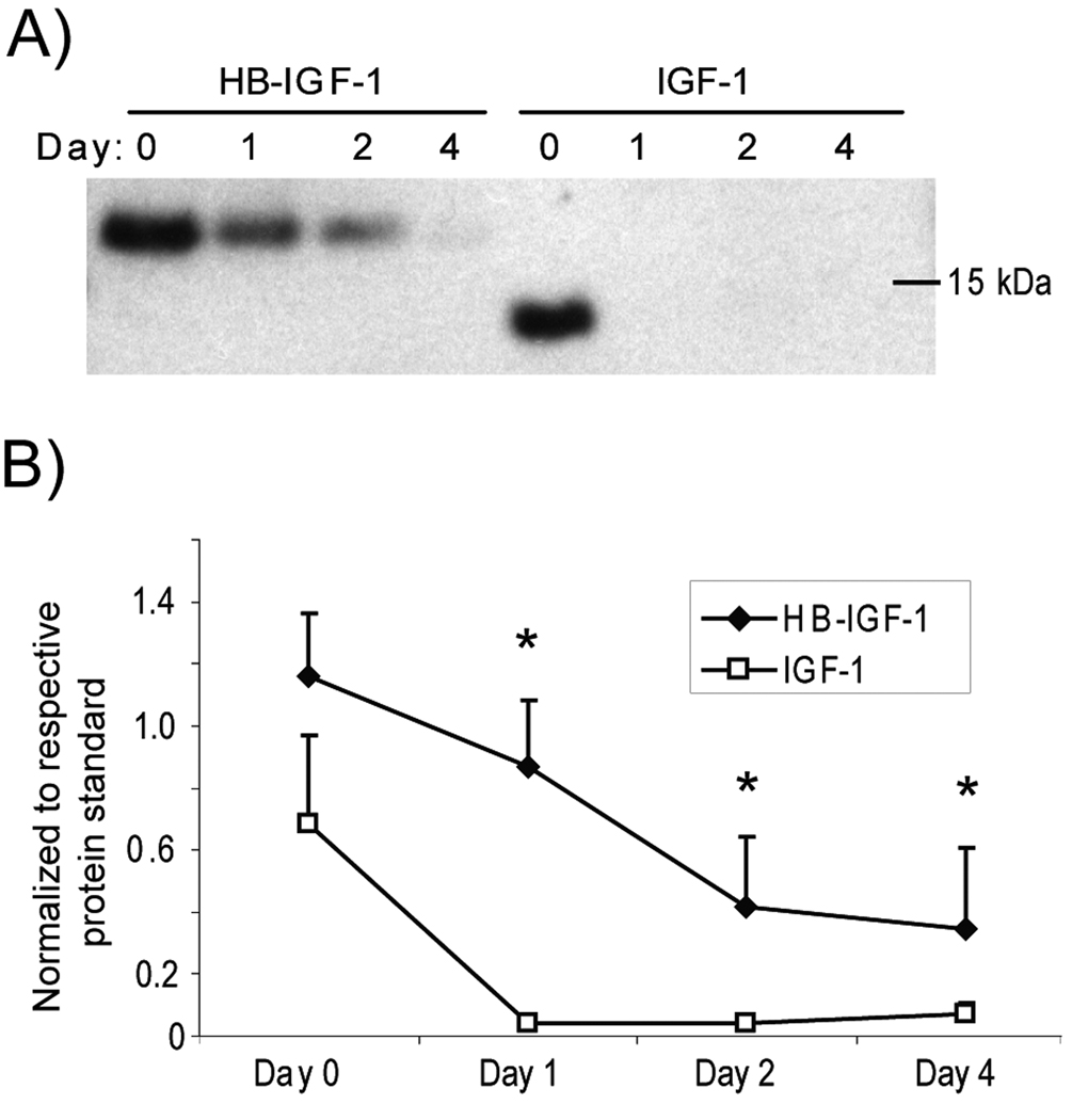
Objective: Insulin-like growth factor 1 (IGF-1) stimulates cartilage repair but is not a practical therapy due to its short half-life. We have previously modified IGF-1 by adding a heparin-binding domain and have shown that this fusion protein (HB-IGF-1) stimulates sustained proteoglycan synthesis in cartilage. This study was undertaken to examine the mechanism by which HB-IGF-1 is retained in cartilage and to test whether HB-IGF-1 provides sustained growth factor delivery to cartilage in vivo and to human cartilage explants.

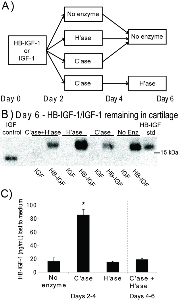
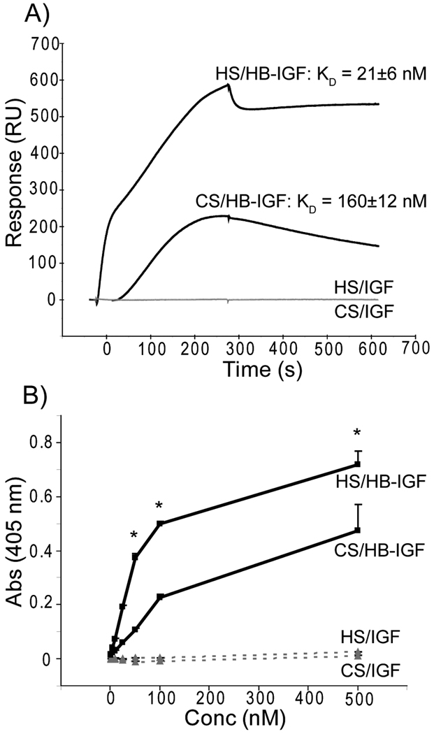
Methods: Retention of HB-IGF-1 and IGF-1 was analyzed by Western blotting. The necessity of heparan sulfate (HS) or chondroitin sulfate (CS) glycosaminoglycans (GAGs) for binding was tested using enzymatic removal and cells with genetic deficiency of HS. Binding affinities of HB-IGF-1 and IGF-1 proteins for isolated GAGs were examined by surface plasmon resonance and enzyme-linked immunosorbent assay.

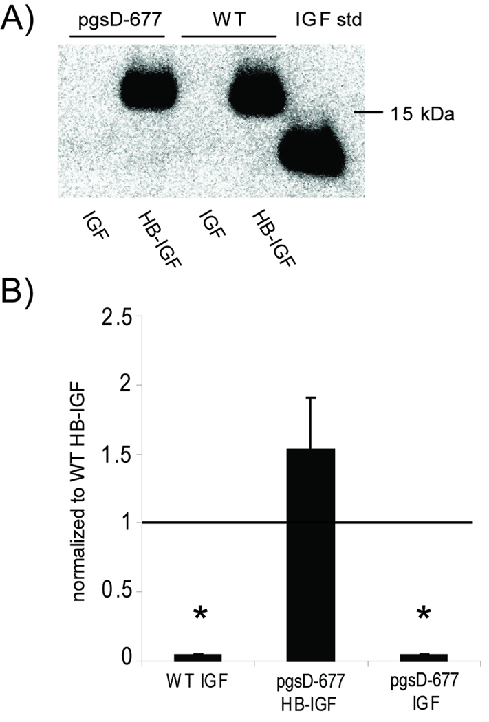
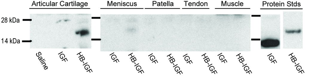
Results: In cartilage explants, chondroitinase treatment decreased binding of HB-IGF-1, whereas heparitinase had no effect. Furthermore, HS was not necessary for HB-IGF-1 retention on cell monolayers. Binding assays showed that HB-IGF-1 bound both CS and HS, whereas IGF-1 did not bind either. After intraarticular injection in rat knees, HB-IGF-1 was retained in articular and meniscal cartilage, but not in tendon, consistent with enhanced delivery to CS-rich cartilage. Finally, HB-IGF-1 was retained in human cartilage explants but IGF-1 was not.

Conclusion: Our findings indicate that after intraarticular injection in rats, HB-IGF-1 is specifically retained in cartilage through its high abundance of CS. Modification of growth factors with heparin-binding domains may be a new strategy for sustained and specific local delivery to cartilage.


Figures

Figure 1
Figure 1
Retention of HB-IGF-1 in bovine cartilage explants following enzymatic digestion of glycosaminoglycans. (A) HB-IGF-1 or IGF-1 was incubated with cartilage disks for two days (Day 0 to Day 2) followed by treatment with no enzyme, chondroitinase (C’ase), or heparitinase (H’ase) for an additional two days (Day 2 to Day 4). At Day 4, a subset of chondroitinase-treated disks was incubated with heparitinase (C’ase+H’ase) for two days while all remaining disks were kept in enzyme-free medium. (B) Western analysis of HB-IGF-1 or IGF-1 remaining in the cartilage tissue at Day 6. The blot shown is representative of four repeats. (C) ELISA of HB-IGF-1 released to the medium following 48-hour enzyme treatment of cartilage explants (Days 2–4 for No enzyme, C’ase, and H’ase conditions; Days 4–6 for C’ase + H’ase). mean ± SEM, * vs. No enzyme, n=4, p<0.001.
Figure 2
Figure 2
Retention of HB-IGF-1 on cells lacking heparan sulfate. (A) HB-IGF-1 (HB-IGF) and IGF-1 (IGF) were incubated with mutant CHO cells unable to produce heparan sulfate (pgsD-677) and wildtype CHO cells (WT), then washed in PBS. Western analysis of the cell lysates for IGF showed that HB-IGF-1 remained bound to cells with or without the presence of heparan sulfate, whereas IGF-1 binding was not detectable. (B) Densitometry of Western blots from four repeated experiments, each normalized to wild-type HB-IGF-1 binding. mean ± SEM, * vs. WT HB, p<0.005.
Figure 3
Figure 3
Binding analysis of HB-IGF-1 and IGF-1 to isolated glycosaminoglycans. (A) Representative sensorgram for Biacore kinetic analysis over HS or CS surfaces using 250 nM HB-IGF-1 or IGF-1. HB-IGF-1 is shown in the top two curves (black) with corresponding equilibrium dissociation constants, KD, determined from a minimum of three concentrations used during three experimental repeats. IGF-1 (bottom two curves, grey) was unable to bind either surface (RU < 10). (B) Sandwich ELISA detecting absorbance at a given HB-IGF-1 (solid line, black) or IGF-1 (dashed line, grey) concentration resulting from binding HS or CS. Representative of two repeats, each with duplicate wells. n=2, mean ± SEM, * vs. CS, p<0.05.
Figure 4
Figure 4
Retention of HB-IGF-1 in vivo. Western blot showing retained IGF-1 (IGF) or HB-IGF-1 (HB-IGF) in rat articular cartilage, meniscus, patella, patellar tendon, or muscle extracts one day after intra-articular injection of IGF-1, HB-IGF-1, or saline.
Figure 5
Figure 5
Retention of HB-IGF-1 in human cartilage explants. Human cartilage (grade 0 to 2) was incubated with HB-IGF-1 or IGF-1 for two days (Day -2 to 0), washed, and incubated in IGF-free medium for 4 days (Days 0–4). (A) Amount of HB-IGF-1 or IGF-1 remaining in cartilage after 0–4 days. The experiment was performed on cartilage from four donors and a representative Western blot is shown. (B) Analysis of four Western blots by densitometry, normalized to the density of 5 ng of the respective protein standard. mean ± SEM, * vs. IGF-1, p<0.05.

References

    1. Goldring MB. Update on the biology of the chondrocyte and new approaches to treating cartilage diseases. Best Practice & Research Clinical Rheumatology. 2006;20:1003–1025. - PubMed
    1. Bonassar LJ, Grodzinsky AJ, Frank EH, Davila SG, Bhaktav NR, Trippel SB. The effect of dynamic compression on the response of articular cartilage to insulin-like growth factor-I. J Orthop Res. 2001;19:11–17. - PubMed
    1. McQuillan DJ, Handley CJ, Campbell MA, Bolis S, Milway VE, Herinton AC. Stimulation of proteoglycan biosynthesis by serum and insulin-like growth factor-I in cultured bovine articular cartilage. Biochem J. 1986;240:423–430. - PMC - PubMed
    1. Curtis AJ, Devenish RJ, Handley CJ. Modulation of aggrecan and link-protein synthesis in articular cartilage. Biochem J. 1992;288:721–726. - PMC - PubMed
    1. Hui W, Rowan AD, Cawston T. Modulation of the expression of matrix metalloproteinase and tissue inhibitors of metalloproteinases by TGF-β1 and IGF-1 in primary human articular and bovine nasal chondrocytes stimulated with TNF-α. Cytokine. 2001;16:31–35. - PubMed

Publication types

MeSH terms