Review
. 2010 Aug;5(8):1185-201.
doi: 10.2217/fmb.10.91.

Recent insights into Pasteurella multocida toxin and other G-protein-modulating bacterial toxins

Affiliations
Review

Recent insights into Pasteurella multocida toxin and other G-protein-modulating bacterial toxins

Brenda A Wilson et al. Future Microbiol. 2010 Aug.

Abstract

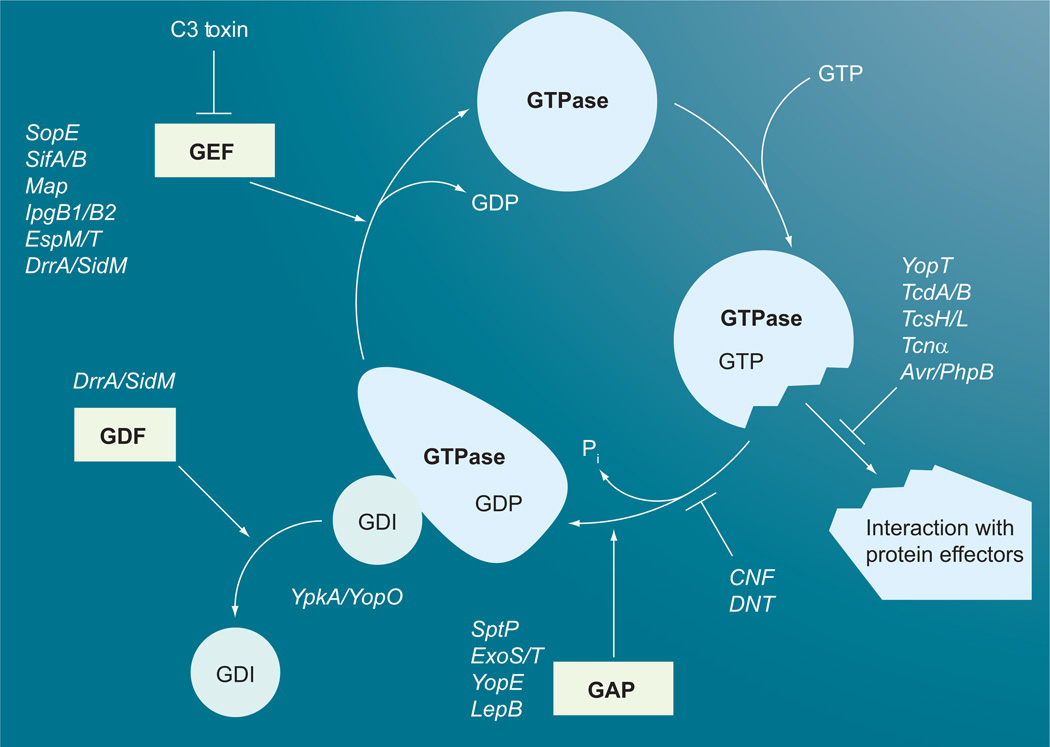
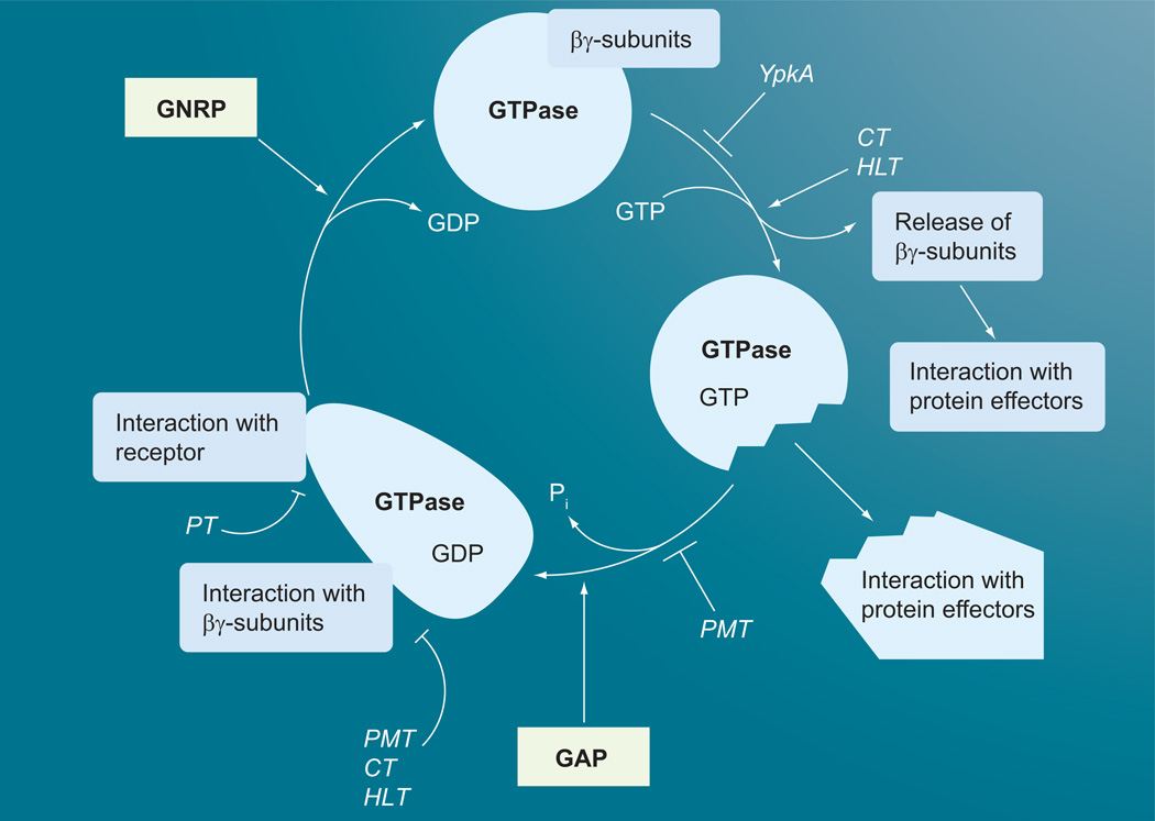
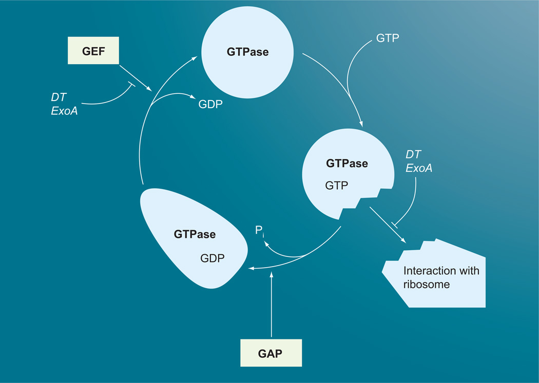
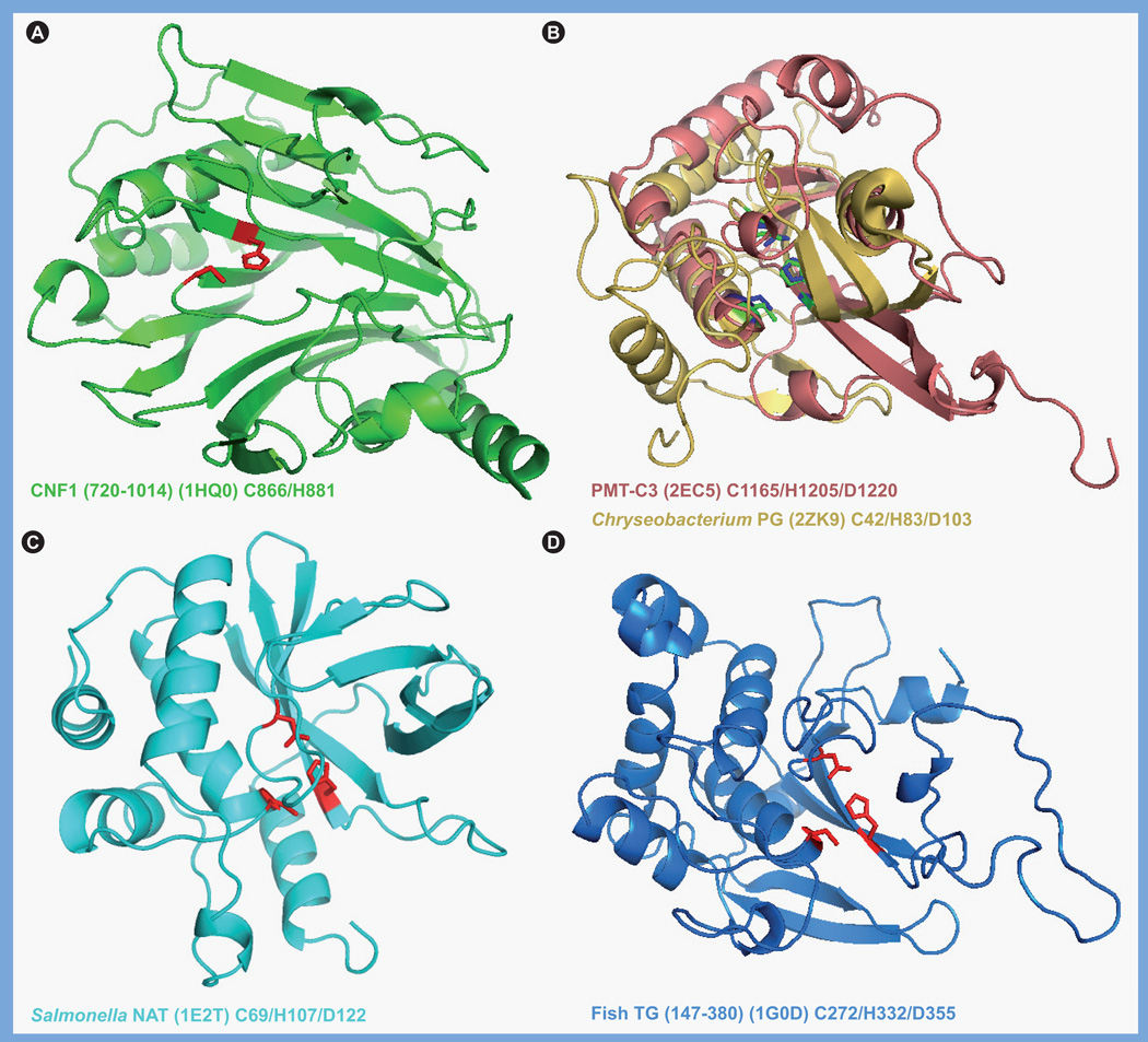
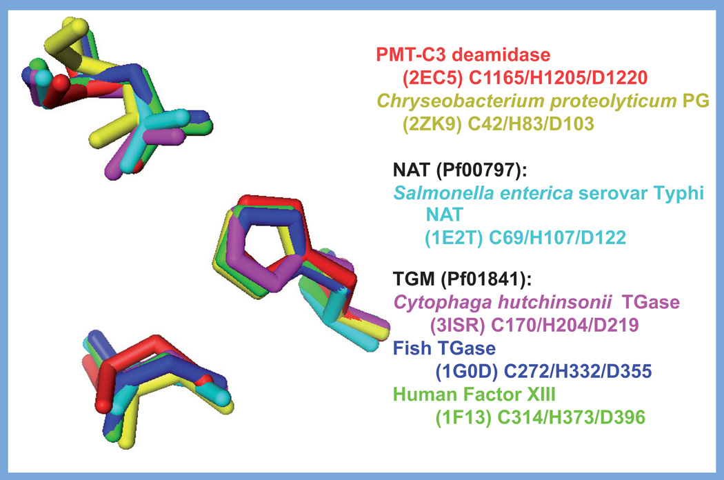
Over the past few decades, our understanding of the bacterial protein toxins that modulate G proteins has advanced tremendously through extensive biochemical and structural analyses. This article provides an updated survey of the various toxins that target G proteins, ending with a focus on recent mechanistic insights in our understanding of the deamidating toxin family. The dermonecrotic toxin from Pasteurella multocida (PMT) was recently added to the list of toxins that disrupt G-protein signal transduction through selective deamidation of their targets. The C3 deamidase domain of PMT has no sequence similarity to the deamidase domains of the dermonecrotic toxins from Escherichia coli (cytotoxic necrotizing factor [CNF]1-3), Yersinia (CNFY) and Bordetella (dermonecrotic toxin). The structure of PMT-C3 belongs to a family of transglutaminase-like proteins, with active site Cys-His-Asp catalytic triads distinct from E. coli CNF1.


Conflict of interest statement

Financial & competing interests disclosure

The authors have no other relevant affiliations or financial involvement with any organization or entity with a financial interest in or financial conflict with the subject matter or materials discussed in the manuscript apart from those disclosed.

No writing assistance was utilized in the production of this manuscript.

Figures

Figure 1
Figure 1. GTPase cycle of small G proteins and points of toxin interactions
Small GTPase binds GDI in the inactive GDP-bound form. GEF facilitates the release of GDI and GDP and the GTPase then binds GTP. The active GTP-bound form interacts with its downstream effectors. Subsequent interaction with GAP stimulates the hydrolysis of GTP to GDP, which converts the GTPase back into its inactive form. Large clostridial toxins (TcdA, TcdB, TcsH, TcsL and Tcnα), YopT and Avr/PhpB interfere with the GTPase interaction with effectors. CNF and DNT block GTPase activity, while the type III secretion system (T3SS) effectors SptP, ExoS, ExoT and YopE, and the T4SS effector LepB, act as GAPs to stimulate GTP hydrolysis. The T3SS effector YpkA/YopO acts as a GDI to prevent release of GDP. The clostridial C3 toxin blocks GEF interaction with the G protein, while the T3SS effectors SopE, SifA, SifB, Map, EspM, EspT, IpgB1 and lpgB2 act as GEFs. The T4SS effector DrrA/SidM acts as both a GEF and a GDF. CNF: Cytotoxic necrotizing factor; DNT: Dermonecrotic toxin; GAP: GTPase activating protein; GDI: Guanine nucleotide dissociation inhibitor; GEF: Guanine nucleotide exchange factor.
Figure 2
Figure 2. GTPase cycle of heterotrimeric G proteins and points of toxin interactions
Heterotrimeric GTPase α subunit binds βγ subunits in the inactive GDP-bound form. The G-protein-coupled receptor bound to its ligand acts as GNRP to stimulate the release of GDP and the α subunit then binds GTP and the βγ subunits dissociate to interact with their downstream effectors. The dissociated active GTP-bound α subunit then interacts with its downstream effectors. PMT, CT and HLT lock the α subunit in its active form and prevent interaction with the βγ subunits and the receptor. PT locks the G protein in its heterotrimeric inactive form and prevents its interaction with the receptor. YpkA prevents GDP/GTP binding to the α subunit. CT: Cholera toxin; GNRP: Guanine nucleotide release protein; HLT: Heat-labile enterotoxins; PMT: Pasteurella multocida toxin; PT: Pertussis toxin.
Figure 3
Figure 3. GTPase cycle of large G proteins and points of toxin interactions
ADP ribosylation of elongation factor (EF)-2 by DT or ExoA blocks interaction of GTP-bound EF-2 with the ribosome in the pretranslocation phase of the peptideelongation cycle, which prevents formation of the high-affinity complex and stimulation of the GAP activity of EF-2 by the ribosome. ADP ribosylation of EF-2 does not interfere with GTP or GDP binding to EF-2, but may inhibit the exchange of GDP with GTP. DT: Diphtheria toxin; GAP: GTPase activating protein; GEF: Guanine nucleotide exchange factor.
Figure 4
Figure 4. Structures of toxin-like deamidase/transglutaminase domains
Shown are the structural folds of the catalytic domains of representative members of the cytotoxic necrotizing factor/dermonecrotic toxin-like family and the PMT-like family of deamidases/ TGases, with the respective active site His–Cys dyad or His–Cys–Asp triad indicated. (A) Catalytic domain of CNF1 (PDB 1HQ0), residues 720–1014 shown in green, with the Cys-866 and His-881 shown in red. (B) Superimposition of the catalytic domains of PMT (PDB 2EC5), residues 1105–1285 shown in pink, with Cys-1165, His-1205 and Asp-1220 shown in blue, and the protein glutaminase from Chryseobacterium proteolyticum (PDB 2ZK9), shown in yellow, with Cys-42, His-83, and Asp-103 shown in green. (C) Catalytic domain of the arylamine N-acetyltransferase from Salmonella enterica serovar Typhimurium (PDB 1E2T), residues 1–197 shown in cyan, with the Cys-69, His-107, and Asp-122 shown in red. (D) Catalytic domain of the fish-derived TGase from red sea bream (PDB 1G0D), residues 147–380 shown in blue, with Cys-272, His-332 and Asp-355 shown in red. Images were generated with PyMOL using the indicated PDB data files. CNF: Cytotoxic necrotizing factor; NAT: N-acetyltransferase; PDB: Protein Data Bank; PMT: Pasteurella multocida toxin; TGase: Transglutaminase.
Figure 5
Figure 5. Comparison of the active-site catalytic triads of the Pasteurella multocida toxin-like deamidases/transglutaminases
Superimposed images of the active site Cys, His and Asp side chains of the catalytic triads from: PMT-C3 (PDB 2EC5) in red, Chryseobacterium protein glutatminase (PDB 2ZK9) in yellow, Salmonella NAT (PDB 1E2T) in light blue, Cytophaga hutchlnsonll TGase (PDB 3ISR) in magenta, fish-derived TGase (PDB 1G0D) in purple and human factor XIII (PDB 1F13) in green. Images were generated with PyMOL using the indicated PDB data files. NAT: N-acetyltransferase; PDB: Protein Data Bank; PMT: Pasteurella multocida toxin; TGase: Transglutaminases;

References

    1. Dautin N, Karimova G, Ladant D. Bordetella pertussis adenylate cyclase toxin: a versatile screening tool. Toxicon. 2002;40(10):1383–1387. - PubMed
    1. Fabbri A, Travaglione S, Falzano L, Fiorentini C. Bacterial protein toxins: current and potential clinical use. Curr. Med. Chem. 2008;15(11):1116–1125. - PubMed
    1. Kostrzewa RM, Segura-Aguilar J. Botulinum neurotoxin: evolution from poison, to research tool - onto medicinal therapeutic and future pharmaceutical panacea. Neurotox Res. 2007;12(4):275–290. ▪ Reviews the potential biomedical applications of bacterial toxins.

    1. Lemonnier M, Landraud L, Lemichez E. Rho GTPase-activating bacterial toxins: from bacterial virulence regulation to eukaryotic cell biology. FEMS Microbiol. Rev. 2007;31(5):515–534. - PubMed
    1. Mathew M, Verma RS. Humanized immunotoxins: a new generation of immunotoxins for targeted cancer therapy. Cancer Sci. 2009;100(8):1359–1365. - PMC - PubMed

Publication types

MeSH terms