Review
. 2010 Sep;32(6):881-93.
doi: 10.1111/j.1460-9568.2010.07354.x. Epub 2010 Aug 16.

Functional subdivision of feline spinal interneurons in reflex pathways from group Ib and II muscle afferents; an update

Affiliations
Review

Functional subdivision of feline spinal interneurons in reflex pathways from group Ib and II muscle afferents; an update

Elzbieta Jankowska et al. Eur J Neurosci. 2010 Sep.

Abstract

A first step towards understanding the operation of a neural network is identification of the populations of neurons that contribute to it. Our aim here is to reassess the basis for subdivision of adult mammalian spinal interneurons that mediate reflex actions from tendon organs (group Ib afferents) and muscle spindle secondary endings (group II afferents) into separate populations. Re-examining the existing experimental data, we find no compelling reasons to consider intermediate zone interneurons with input from group Ib afferents to be distinct from those co-excited by group II afferents. Similar patterns of distributed input have been found in subpopulations that project ipsilaterally, contralaterally or bilaterally, and in both excitatory and inhibitory interneurons; differences in input from group I and II afferents to individual interneurons showed intra- rather than inter-population variation. Patterns of reflex actions evoked from group Ib and II afferents and task-dependent changes in these actions, e.g. during locomotion, may likewise be compatible with mediation by premotor interneurons integrating information from both group I and II afferents. Pathological changes after injuries of the central nervous system in humans and the lineage of different subclasses of embryonic interneurons may therefore be analyzed without need to consider subdivision of adult intermediate zone interneurons into subpopulations with group Ib or group II input. We propose renaming these neurons 'group I/II interneurons'.


Figures

Figure 1
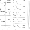
Figure 1. Examples of different degrees of contribution of group I and group II afferents to excitation of intermediate zone interneurons following stimulation of muscle nerves
Each panel shows recordings from a different interneuron (upper traces) and from the surface of the spinal cord close to the dorsal root entry zone (lower traces). In A–C are extracellular recordings from three neurons activated by stimulation of group I afferents (A), both group I and II afferents (B) or only group II afferents, respectively. They were activated by stimulation of the gastrocnemius-soleus (GS) nerve at 1.8 times threshold and stimulation of the quadriceps (Q) nerve at 5 times threshold as indicated. D–H show intracellular recordings from 5 interneurons. Monosynaptic EPSPs evoked in these interneurons from the quadriceps nerve ranged from group I alone (D) to group II alone (H), but with different combinations in between, as schematically indicated to the right. Dotted lines indicate onset times of the spikes and of the earliest EPSPs from group I and group II afferents, at latencies of 0.7–0.9 ms and 1.7–1.9 ms from the afferent volleys respectively. Negativity is downwards in intracellular records and upwards in extracellular records. Rectangular pulses at the beginning of the records are calibration pulses (0.2 mV).
Figure 2
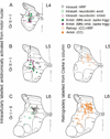
Figure 2. Locations of intermediate zone interneurons with input from group I and/or group II afferents
Location of the different samples of intermediate zone interneurons indicated on Rexed´s diagrams of the L4, L5 and L6 segments. A – C, Open circles, locations of interneurons labelled intracellularly with HRP [A, data from Fig. 1 in (Bras et al., 1989a) and from Fig. 11 in (Bannatyne et al., 2009); B and C, data from Fig. 10 in (Jankowska et al., 1981a and Fig. 1 and 2 in Czarkowska et al., 1981)] or a mixture of rhodamine dextran and neurobiotin [data from Fig. 5 in (Bannatyne et al., 2009)]. Green, glutamatergic (excitatory) interneurons; red, glycinergic (inhibitory) interneurons; black, interneurons with undefined transmitter phenotype. Most of these neurons were antidromically activated by stimuli applied in ipsilateral GS or HS motor nuclei (MN) in the L7 segment. Filled circles, antidromically activated, but extracellularly recorded interneurons that evoked population EPSPs (green) or IPSPs (red) in hindlimb motorneurons as found by spike triggered averaging [from Fig. 5 in (Cavallari et al., 1987)]. D and E, Open circles, location of interneurons labelled by retrograde transport of HRP from Clarke´s column (CC) [from Fig. 6 in (Hongo et al., 1983a)]; filled circles, location of interneurons antidromically activated from Clarke´s column [from Fig. 7 in (Hongo et al., 1983a)].
Figure 3
Figure 3. Relationships between the transmitter phenotype, location, projection areas and input to interneurons activated by muscle afferents
A, Examples of excitatory and inhibitory interneurons located in the dorsal horn (top), the intermediate zone (middle) and lamina VIII (bottom), with their typical terminal projection areas (shaded). B, Diagrams summarizing the axonal projections for the groups of neurons at these locations that we have studied: glutamatergic and glycinergic. The circles represent cell bodies of the interneurons (all located to the left of the midline indicated by the dotted line, crosses in the circles represent contralaterally–projecting neurons) while rectangular boxes to the left and right of these circles represent ipsilateral (i) and contralateral (co) projection areas within the dorsal horn, intermediate zone and the ventral horn (including motor nuclei) to which they project. Green and red boxes denote regions of axonal projections of excitatory and inhibitory interneurons respectively. Black boxes denote regions in which no projections from these interneurons were found. Note that all intermediate zone interneurons projecting to the motor nuclei had also terminal projection areas that were outside motor nuclei, showing that they target other neurons as well as motoneurons. C, the main sources of input to these interneurons. Modified from Figs. 6, 7 and 11 in Bannatyne et al. (2009), Figs. 7 and 8 in Bannatyne et al. (2006) and Figs. 5 and 9 in Bannatyne et al. (2003).
Figure 4
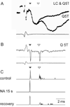
Figure 4. Examples of differential modulation of synaptic actions of group I and II afferents on motoneurons and intermediate zone interneurons
Responses in a motoneuron (A) and an intermediate zone interneuron (B & C) both of which were evoked by both group I and group II afferents (filled and open arrowheads indicate the early effects of group I and later effects of II afferents respectively). A, Intracellular records from a GS motoneuron, illustrating the effects of stimuli applied in the region of origin of the descending noradrenergic neurons, in Locus coeruleus/subcoeruleus (LC) on IPSPs evoked by stimulation of both group I and II afferents in the quadriceps (Q); lower trace, afferent volley from the cord dorsum. Note that control records displayed both an early (group I,) and a late (group II) component: the late component was very substantially suppressed when Q stimulation was preceded by LC conditioning stimulation (at a conditioning-testing interval of 160 ms). Modified from Jankowska et al. (1993a). B, Records from an intermediate zone interneuron responding with an early (group I) and a later (group II) spikes to the same intensity of Q stimulation and record of afferent volleys from the cord dorsum. C, peristimulus time histograms of spike potentials (exceeding the discrimination level indicated by the dotted line) for the interneuron illustrated in B. They show responses evoked by 20 consecutive stimuli before, during and after ionophoretic ejection of NA from a second micropipette positioned close to the same interneuron. Note that the late responses disappeared during application of NA while the early responses persisted. Modified from Fig. 5 in Jankowska et al.(2000).
Figure 5
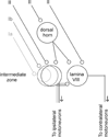
Figure 5. Simplifying diagram of relationships between intermediate zone interneurons co-excited by group I and II afferents and dorsal horn and laminaVIII interneurons with input from group I and II afferents
The diagram takes only into account the distribution of the excitatory input to these neurons. However interactions have been found both between excitatory and inhibitory interneurons with input from group Ib and II afferents.

References

    1. Aggelopoulos NC, Bawa P, Edgley SA. Activation of midlumbar neurones by afferents from anterior hindlimb muscles in the cat. J. Physiol. (Lond) 1996a;497:795–802. - PMC - PubMed
    1. Aggelopoulos NC, Burton MJ, Clarke RW, Edgley SA. Characterization of a descending system that enables crossed group II inhibitory reflex pathways in the cat spinal cord. J. Neurosci. 1996b;16:723–729. - PMC - PubMed
    1. Al-Mosawie A, Wilson JM, Brownstone RM. Heterogeneity of V2-derived interneurons in the adult mouse spinal cord. Eur. J. Neurosci. 2007;26:3003–3015. - PubMed
    1. Alstermark B, Kummel H. Transneuronal transport of wheat germ agglutinin conjugated horseradish peroxidase into last order spinal interneurones projecting to acromio- and spinodeltoideus motoneurones in the cat. 1. Location of labelled interneurones and influence of synaptic activity on the transneuronal transport. Exp. Brain Res. 1990;80:83–95. - PubMed
    1. Alvarez FJ, Fyffe RE. The continuing case for the Renshaw cell. J. Physiol. (Lond) 2007;584:31–45. - PMC - PubMed

Publication types