. 2011 Jan;132(1):39-48.
doi: 10.1111/j.1365-2567.2010.03335.x. Epub 2010 Aug 17.

Osteopontin is up-regulated and associated with neutrophil and macrophage infiltration in glioblastoma

Affiliations

Osteopontin is up-regulated and associated with neutrophil and macrophage infiltration in glioblastoma

Nadia A Atai et al. Immunology. 2011 Jan.

Abstract

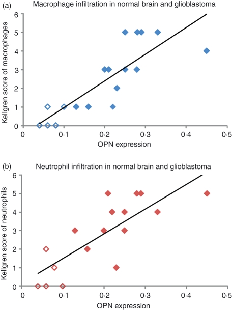
Osteopontin (OPN) is a glycophosphoprotein with multiple intracellular and extracellular functions. In vitro, OPN enhances migration of mouse neutrophils and macrophages. In cancer, extracellular OPN facilitates migration of cancer cells via its RGD sequence. The present study was designed to investigate whether osteopontin is responsible for neutrophil and macrophage infiltration in human cancer and in particular in glioblastoma. We found that in vitro mouse neutrophil migration was RGD-dependent. In silico, we found that the OPN gene was one of the 5% most highly expressed genes in 20 out of 35 cancer microarray data sets in comparison with normal tissue in at least 30% of cancer patients. In some types of cancer, such as ovarian cancer, lung cancer and melanoma, the OPN gene was one of those with the highest expression levels in at least 90% of cancer patients. In glioblastoma, the most invasive type of brain tumours/glioma, but not in lower grades of glioma it was one of the 5% highest expressed genes in 90% of patients. In situ, we found increased protein levels of OPN in human glioblastoma versus normal human brain confirming in silico results. OPN protein expression was co-localized with neutrophils and macrophages. In conclusion, OPN in tumours not only induces migration of cancer cells but also of leucocytes.


Figures

Figure 1
Figure 1
Quantitative analysis (log10 scale) of in vitro recruitment of wild-type (WT) and osteopontin-deficient (OPN−/−) neutrophils by three types of exogenous OPN [macrophage OPN (mϕ-OPN), recombinant OPN (rec-OPN) and mutated OPN (mut-OPN)] and representative images of recruited neutrophils by each type of OPN. Differences between the effects of the three types of OPN and between WT and OPN−/− neutrophils are significant (P < 0·05). Data are presented as log10 values because this results in a normal distribution of the data in all cases.
Figure 2
Figure 2
In silico analysis of expression of the osteopontin gene OPN in cancer data sets. Heatmap of percentile fold change and percentage over-expression of the OPN gene expression in 20 cancer data sets that have a percentile fold change > 0·95 and percentage over-expression > 30% meaning that the OPN gene is one of the 5% of genes that are most highly expressed in cancer in at least 30% of the patients. Number of normal tissue samples (N) and cancer samples (C) per data set are given.
Figure 3
Figure 3
Percentile fold change and percentage over-expression of expression of osteopontin gene OPN in different WHO grades and histological grades of glioma. (a, c) Heatmap of WHO grade (WHO-2, WHO-3 and WHO-4 = glioblastoma) in the data sets of Bredel et al. and Kotliarov et al. The number of tumour samples included are nine WHO-2, 10 WHO-3 and 31 WHO-4 grades in the Bredel et al. data set and 45 WHO-2, 31 WHO-3 and 81 WHO-4 grades in the Kotliarov et al. data set. (b, d) Heatmap of histological grades (oligodenroglial, astrocytic and glioblastoma) in the data sets of Bredel et al. and Kotliarov et al. The number of tumour samples included are nine oligodenroglial, two astrocytic and 31 glioblastoma in the Bredel et al. data set and 50 oligodenroglial, 26 astrocytic and 81 glioblastoma in the Kotliarov et al. data set.
Figure 4
Figure 4
Immunohistochemical analysis of osteopontin (OPN) protein expression (a), neutrophils (b) and macrophages (c) in normal human brain tissue. OPN is present in endothelial cells (arrows) and microglial cells (arrowheads). Neutrophils and macrophages are absent. Bar = 40 μm.
Figure 6
Figure 6
Relationship between quantitative image analysis data of osteopontin (OPN) protein expression in situ and the semi-quantative scores of macrophages (a; P < 0·05) and neutrophils (b; P < 0·05) in non-cancerous human brain tissue (n = 5) and glioblastoma (n = 13).
Figure 5
Figure 5
Immunohistochemical analysis of osteopontin (OPN) protein expression (a, d, g), neutrophils (b, e, h) and macrophages (c, f, i) in human glioblastoma samples. The OPN is highly expressed extracellularly (CC) around necrotic areas (N) and blood vessels (BV), where neutrophils and macrophages are present. Bars represent 40 μm (a, b, c, g, h, i) and 20 μm (d, e, f).

Similar articles

Cited by

References

    1. Sodek J, Ganss B, Mckee MD. Osteopontin. Crit Rev Oral Biol Med. 2000;11:279–303. - PubMed
    1. Sodek J, Zhu B, Huynth MH, Brown TJ, Ringuette M. Novel functions of the matricellular proteins osteopontin and osteonectin/SPARC. Connect Tissue Res. 2002;43:308–19. - PubMed
    1. Denhardt DT, Giachelli CM, Rittling SR. Role of osteopontin in cellular signaling and toxicant injury. Annu Rev Pharmacol Toxicol. 2001;41:723–49. - PubMed
    1. Wai PY, Kuo PC. Osteopontin: regulation in tumor metastasis. Cancer Metastasis Rev. 2008;27:103–18. - PubMed
    1. Ramaiah SK, Rittling S. Pathophysiological role of osteopontin in hepatic inflammation, toxicity, and cancer. Toxicol Sci. 2008;103:4–13. - PubMed

LinkOut - more resources