Primary pneumonic plague in the African Green monkey as a model for treatment efficacy evaluation
- PMID: 20722770
- PMCID: PMC2991483
- DOI: 10.1111/j.1600-0684.2010.00443.x
Primary pneumonic plague in the African Green monkey as a model for treatment efficacy evaluation
Abstract
Background: Primary pneumonic plague is rare among humans, but treatment efficacy may be tested in appropriate animal models under the FDA 'Animal Rule'.
Methods: Ten African Green monkeys (AGMs) inhaled 44-255 LD(50) doses of aerosolized Yersinia pestis strain CO92. Continuous telemetry, arterial blood gases, chest radiography, blood culture, and clinical pathology monitored disease progression.
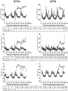
Results: Onset of fever, >39°C detected by continuous telemetry, 52-80 hours post-exposure was the first sign of systemic disease and provides a distinct signal for treatment initiation. Secondary endpoints of disease severity include tachypnea measured by telemetry, bacteremia, extent of pneumonia imaged by chest x-ray, and serum lactate dehydrogenase enzyme levels.
Conclusions: Inhaled Y. pestis in the AGM results in a rapidly progressive and uniformly fatal disease with fever and multifocal pneumonia, serving as a rigorous test model for antibiotic efficacy studies.
© 2010 John Wiley & Sons A/S.
Figures


Similar articles
-
The African Green Monkey Model of Pneumonic Plague and US Food and Drug Administration Approval of Antimicrobials Under the Animal Rule.Clin Infect Dis. 2020 May 21;70(70 Suppl 1):S51-S59. doi: 10.1093/cid/ciz1233. Clin Infect Dis. 2020. PMID: 32435803 Free PMC article.
-
Levofloxacin cures experimental pneumonic plague in African green monkeys.PLoS Negl Trop Dis. 2011 Feb 8;5(2):e959. doi: 10.1371/journal.pntd.0000959. PLoS Negl Trop Dis. 2011. PMID: 21347450 Free PMC article.
-
Milestones in progression of primary pneumonic plague in cynomolgus macaques.Infect Immun. 2010 Jul;78(7):2946-55. doi: 10.1128/IAI.01296-09. Epub 2010 Apr 12. Infect Immun. 2010. PMID: 20385751 Free PMC article.
-
Pneumonic Plague: The Darker Side of Yersinia pestis.Trends Microbiol. 2016 Mar;24(3):190-197. doi: 10.1016/j.tim.2015.11.008. Epub 2015 Dec 14. Trends Microbiol. 2016. PMID: 26698952 Review.
-
Imaging analysis of pneumonic plague infection in Xizang, China: a case report and literature review.BMC Pulm Med. 2024 Aug 1;24(1):378. doi: 10.1186/s12890-024-03187-3. BMC Pulm Med. 2024. PMID: 39090583 Free PMC article. Review.
Cited by
-
The African Green Monkey Model of Pneumonic Plague and US Food and Drug Administration Approval of Antimicrobials Under the Animal Rule.Clin Infect Dis. 2020 May 21;70(70 Suppl 1):S51-S59. doi: 10.1093/cid/ciz1233. Clin Infect Dis. 2020. PMID: 32435803 Free PMC article.
-
Model systems to study plague pathogenesis and develop new therapeutics.Front Microbiol. 2010 Nov 4;1:119. doi: 10.3389/fmicb.2010.00119. eCollection 2010. Front Microbiol. 2010. PMID: 21687720 Free PMC article.
-
Peripheral Blood Biomarkers of Disease Outcome in a Monkey Model of Rift Valley Fever Encephalitis.J Virol. 2018 Jan 17;92(3):e01662-17. doi: 10.1128/JVI.01662-17. Print 2018 Feb 1. J Virol. 2018. PMID: 29118127 Free PMC article.
-
Prevention of pneumonic plague in mice, rats, guinea pigs and non-human primates with clinical grade rV10, rV10-2 or F1-V vaccines.Vaccine. 2011 Sep 2;29(38):6572-83. doi: 10.1016/j.vaccine.2011.06.119. Epub 2011 Jul 16. Vaccine. 2011. PMID: 21763383 Free PMC article.
-
Levofloxacin cures experimental pneumonic plague in African green monkeys.PLoS Negl Trop Dis. 2011 Feb 8;5(2):e959. doi: 10.1371/journal.pntd.0000959. PLoS Negl Trop Dis. 2011. PMID: 21347450 Free PMC article.
References
-
- Boles JW, Pitt ML, LeClaire RD, Gibbs PH, Ulrich RG, Bavari S. Correlation of Body Temperature with Protection against Staphylococcal Enterotoxin B Exposure and Use in Determining Vaccine Dose-Schedule. Vaccine. 2003;21:2791–2796. - PubMed
-
- Boulanger LL, Ettestad P, Fogarty JD, Dennis DT, Romig D, Mertz G. Gentamicin and Tetracyclines for the Treatment of Human Plague: Review of 75 Cases in New Mexico, 1985-1999. Clin Infect Dis. 2004;38:663–669. - PubMed
-
- Burmeister RW, Tigertt WD, Overholt EL. Laboratory-Acquired Pneumonic Plague. Report of a Case and Review of Previous Cases. Ann Intern Med. 1962;56:789–800. - PubMed
Publication types
MeSH terms
Substances
Grants and funding
LinkOut - more resources
Full Text Sources
Medical
Research Materials

