

Unravelling the development of the visual cortex: implications for plasticity and repair
- PMID: 20722872
- PMCID: PMC2992420
- DOI: 10.1111/j.1469-7580.2010.01275.x
Unravelling the development of the visual cortex: implications for plasticity and repair
Abstract
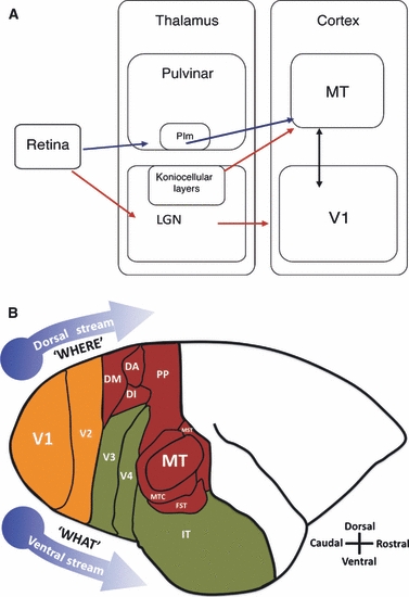
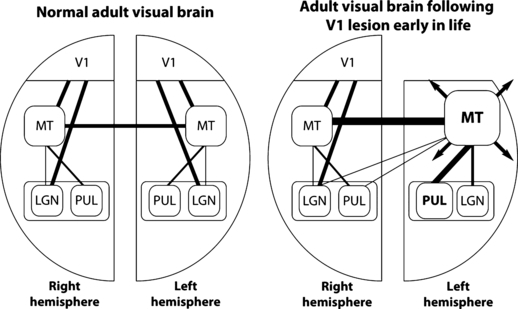
The visual cortex comprises over 50 areas in the human, each with a specified role and distinct physiology, connectivity and cellular morphology. How these individual areas emerge during development still remains something of a mystery and, although much attention has been paid to the initial stages of the development of the visual cortex, especially its lamination, very little is known about the mechanisms responsible for the arealization and functional organization of this region of the brain. In recent years we have started to discover that it is the interplay of intrinsic (molecular) and extrinsic (afferent connections) cues that are responsible for the maturation of individual areas, and that there is a spatiotemporal sequence in the maturation of the primary visual cortex (striate cortex, V1) and the multiple extrastriate/association areas. Studies in both humans and non-human primates have started to highlight the specific neural underpinnings responsible for the maturation of the visual cortex, and how experience-dependent plasticity and perturbations to the visual system can impact upon its normal development. Furthermore, damage to specific nuclei of the visual cortex, such as the primary visual cortex (V1), is a common occurrence as a result of a stroke, neurotrauma, disease or hypoxia in both neonates and adults alike. However, the consequences of a focal injury differ between the immature and adult brain, with the immature brain demonstrating a higher level of functional resilience. With better techniques for examining specific molecular and connectional changes, we are now starting to uncover the mechanisms responsible for the increased neural plasticity that leads to significant recovery following injury during this early phase of life. Further advances in our understanding of postnatal development/maturation and plasticity observed during early life could offer new strategies to improve outcomes by recapitulating aspects of the developmental program in the adult brain.
© 2010 The Author. Journal of Anatomy © 2010 Anatomical Society of Great Britain and Ireland.
Figures
Similar articles
-
Contribution of thalamic input to the specification of cytoarchitectonic cortical fields in the primate: effects of bilateral enucleation in the fetal monkey on the boundaries, dimensions, and gyrification of striate and extrastriate cortex.J Comp Neurol. 1996 Mar 25;367(1):70-89. doi: 10.1002/(SICI)1096-9861(19960325)367:1<70::AID-CNE6>3.0.CO;2-G. J Comp Neurol. 1996. PMID: 8867284
-
Cross-modal plasticity after monocular enucleation of the adult rabbit.Exp Brain Res. 2002 Jun;144(4):423-9. doi: 10.1007/s00221-002-1087-8. Epub 2002 Apr 17. Exp Brain Res. 2002. PMID: 12037628
-
Development and plasticity of the primary visual cortex.Neuron. 2012 Jul 26;75(2):230-49. doi: 10.1016/j.neuron.2012.06.009. Neuron. 2012. PMID: 22841309 Free PMC article. Review.
-
Circuitry Underlying Experience-Dependent Plasticity in the Mouse Visual System.Neuron. 2020 Apr 8;106(1):21-36. doi: 10.1016/j.neuron.2020.01.031. Neuron. 2020. PMID: 32272065 Free PMC article. Review.
-
Role of afferent activity in the development of cortical specification.Results Probl Cell Differ. 2002;39:139-56. doi: 10.1007/978-3-540-46006-0_7. Results Probl Cell Differ. 2002. PMID: 12353467 Review.
Cited by
-
Longitudinal development of cortical and subcortical gray matter from birth to 2 years.Cereb Cortex. 2012 Nov;22(11):2478-85. doi: 10.1093/cercor/bhr327. Epub 2011 Nov 22. Cereb Cortex. 2012. PMID: 22109543 Free PMC article.
-
Thalamocortical circuits for the formation of hierarchical pathways in the mammalian visual cortex.Front Neural Circuits. 2023 Apr 17;17:1155195. doi: 10.3389/fncir.2023.1155195. eCollection 2023. Front Neural Circuits. 2023. PMID: 37139079 Free PMC article. Review.
-
Renewed focus on the developing human neocortex.J Anat. 2010 Oct;217(4):276-88. doi: 10.1111/j.1469-7580.2010.01281.x. J Anat. 2010. PMID: 20979582 Free PMC article. Review.
-
Cross-sectional and longitudinal changes in category selectivity in visual cortex following pediatric cortical resection.Commun Biol. 2025 Aug 12;8(1):1200. doi: 10.1038/s42003-025-08554-2. Commun Biol. 2025. PMID: 40797085 Free PMC article.
-
Differential functional reorganization of ventral and dorsal visual pathways following childhood hemispherectomy.bioRxiv [Preprint]. 2023 Aug 5:2023.08.03.551494. doi: 10.1101/2023.08.03.551494. bioRxiv. 2023. Update in: Dev Cogn Neurosci. 2023 Dec;64:101323. doi: 10.1016/j.dcn.2023.101323. PMID: 37577633 Free PMC article. Updated. Preprint.
References
-
- Adams MM, Hof PR, Gattass R, et al. Visual cortical projections and chemoarchitecture of macaque monkey pulvinar. J Comp Neurol. 2000;419:377–393. - PubMed
-
- Ang LC, Munoz DG, Shul D, et al. SMI-32 immunoreactivity in human striate cortex during postnatal development. Brain Res Dev Brain Res. 1991;61:103–109. - PubMed
-
- Angelucci A, Clasca F, Sur M. Anterograde axonal tracing with the subunit B of cholera toxin: a highly sensitive immunohistochemical protocol for revealing fine axonal morphology in adult and neonatal brains. J Neurosci Methods. 1996;65:101–112. - PubMed
-
- Annese J, Gazzaniga MS, Toga AW. Localization of the human cortical visual area MT based on computer aided histological analysis. Cereb Cortex. 2005;15:1044–1053. - PubMed
-
- Atkinson J. Human visual development over the first 6 months of life. A review and a hypothesis. Hum Neurobiol. 1984;3:61–74. - PubMed
Publication types
MeSH terms
Substances
LinkOut - more resources
Full Text Sources
