

Pulmonary hemodynamic responses to in utero ventilation in very immature fetal sheep
- PMID: 20723253
- PMCID: PMC2944277
- DOI: 10.1186/1465-9921-11-111
Pulmonary hemodynamic responses to in utero ventilation in very immature fetal sheep
Abstract
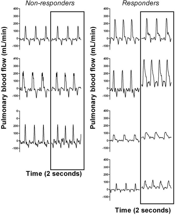
Background: The onset of ventilation at birth decreases pulmonary vascular resistance (PVR) resulting in a large increase in pulmonary blood flow (PBF). As the large cross sectional area of the pulmonary vascular bed develops late in gestation, we have investigated whether the ventilation-induced increase in PBF is reduced in immature lungs.
Methods: Surgery was performed in fetal sheep at 105 d GA (n = 7; term ~147 d) to insert an endotracheal tube, which was connected to a neonatal ventilation circuit, and a transonic flow probe was placed around the left pulmonary artery. At 110 d GA, fetuses (n = 7) were ventilated in utero (IUV) for 12 hrs while continuous measurements of PBF were made, fetuses were allowed to develop in utero for a further 7 days following ventilation.
Results: PBF changes were highly variable between animals, increasing from 12.2 ± 6.6 mL/min to a maximum of 78.1 ± 23.1 mL/min in four fetuses after 10 minutes of ventilation. In the remaining three fetuses, little change in PBF was measured in response to IUV. The increases in PBF measured in responding fetuses were not sustained throughout the ventilation period and by 2 hrs of IUV had returned to pre-IUV control values.
Discussion and conclusion: Ventilation of very immature fetal sheep in utero increased PBF in 57% of fetuses but this increase was not sustained for more than 2 hrs, despite continuing ventilation. Immature lungs can increase PBF during ventilation, however, the present studies show these changes are transient and highly variable.
Figures


Comment in
-
The effects of preterm birth and its antecedents on the cardiovascular system.Acta Obstet Gynecol Scand. 2016 Jun;95(6):652-63. doi: 10.1111/aogs.12880. Epub 2016 Mar 31. Acta Obstet Gynecol Scand. 2016. PMID: 26918772 Review.
Similar articles
-
Increases in lung expansion alter pulmonary hemodynamics in fetal sheep.J Appl Physiol (1985). 2006 Jul;101(1):273-82. doi: 10.1152/japplphysiol.01544.2005. Epub 2006 Mar 30. J Appl Physiol (1985). 2006. PMID: 16575019
-
Positive end-expiratory pressure differentially alters pulmonary hemodynamics and oxygenation in ventilated, very premature lambs.J Appl Physiol (1985). 2005 Oct;99(4):1453-61. doi: 10.1152/japplphysiol.00055.2005. Epub 2005 May 12. J Appl Physiol (1985). 2005. PMID: 15890759
-
Influence of fetal breathing movements on pulmonary hemodynamics in fetal sheep.Pediatr Res. 2004 Dec;56(6):932-8. doi: 10.1203/01.PDR.0000145254.66447.C0. Epub 2004 Oct 6. Pediatr Res. 2004. PMID: 15470203
-
Impact of Acute and Chronic Hypoxia-Ischemia on the Transitional Circulation.Pediatrics. 2021 Mar;147(3):e2020016972. doi: 10.1542/peds.2020-016972. Pediatrics. 2021. PMID: 33622795 Review.
-
The transition from fetal to neonatal circulation: normal responses and implications for infants with heart disease.Semin Perinatol. 1993 Apr;17(2):106-21. Semin Perinatol. 1993. PMID: 8327901 Review.
Cited by
-
Mechanical ventilation induces brainstem inflammation in preterm fetal sheep.Front Pediatr. 2023 Oct 23;11:1225294. doi: 10.3389/fped.2023.1225294. eCollection 2023. Front Pediatr. 2023. PMID: 37936886 Free PMC article.
-
Respiratory Support of the Preterm Neonate: Lessons About Ventilation-Induced Brain Injury From Large Animal Models.Front Neurol. 2020 Aug 14;11:862. doi: 10.3389/fneur.2020.00862. eCollection 2020. Front Neurol. 2020. PMID: 32922358 Free PMC article. Review.
References
-
- Rudolph AM, Heymann MA. Foetal and neonatal physiology Proceedings of the Sir Joseph Barcroft centenary symposium The Physiological Laboratory Cambridge 25 to 27 of july 1972. London: Cambridge University Press; 1973. Control of the Foetal circulation; pp. 89–111.
-
- Dawes GS, MILNE ED, Mott JC, Widdicombe JG. The closure of the foramen ovale after birth. J Physiol. 1953;122:38P. - PubMed
Publication types
MeSH terms
LinkOut - more resources
Full Text Sources
Research Materials
