. 2010 Aug 20;142(4):556-67.
doi: 10.1016/j.cell.2010.07.017.

The monopolin complex crosslinks kinetochore components to regulate chromosome-microtubule attachments

Affiliations

The monopolin complex crosslinks kinetochore components to regulate chromosome-microtubule attachments

Kevin D Corbett et al. Cell. .

Abstract

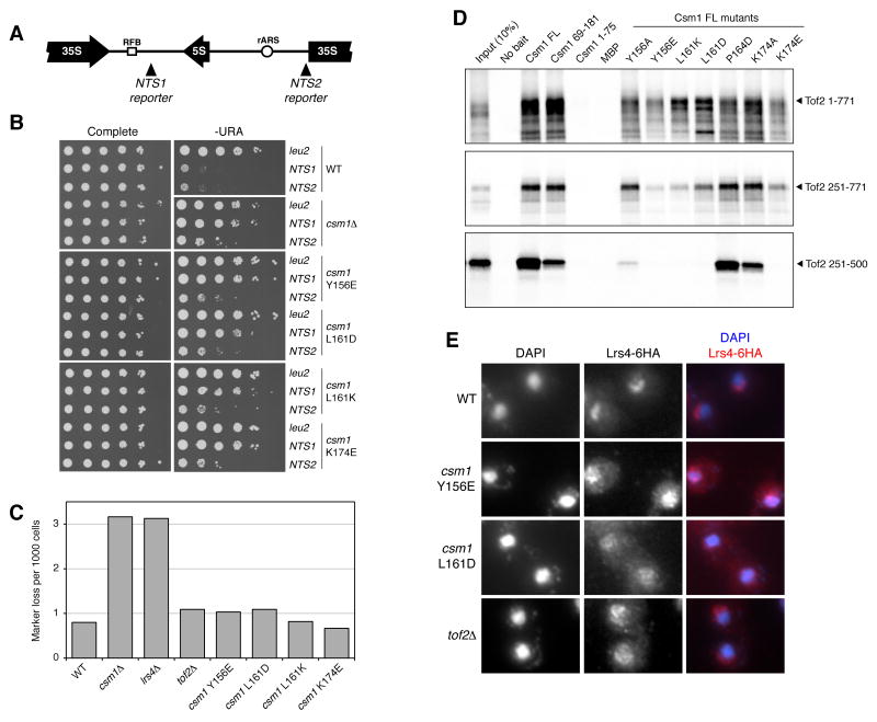
The monopolin complex regulates different types of kinetochore-microtubule attachments in fungi, ensuring sister chromatid co-orientation in Saccharomyces cerevisiae meiosis I and inhibiting merotelic attachment in Schizosaccharomyces pombe mitosis. In addition, the monopolin complex maintains the integrity and silencing of ribosomal DNA (rDNA) repeats in the nucleolus. We show here that the S. cerevisiae Csm1/Lrs4 monopolin subcomplex has a distinctive V-shaped structure, with two pairs of protein-protein interaction domains positioned approximately 10 nm apart. Csm1 presents a conserved hydrophobic surface patch that binds two kinetochore proteins: Dsn1, a subunit of the outer-kinetochore MIND/Mis12 complex, and Mif2/CENP-C. Csm1 point-mutations that disrupt kinetochore-subunit binding also disrupt sister chromatid co-orientation in S. cerevisiae meiosis I. We further show that the same Csm1 point-mutations affect rDNA silencing, probably by disrupting binding to the rDNA-associated protein Tof2. We propose that Csm1/Lrs4 functions as a molecular clamp, crosslinking kinetochore components to enforce sister chromatid co-orientation in S. cerevisiae meiosis I and to suppress merotelic attachment in S. pombe mitosis, and crosslinking rDNA repeats to aid rDNA silencing.


Figures

Figure 1
Figure 1. Structure of Csm1
(A) The Csm1 dimer. One chain is shown in orange (N-terminal coiled-coil, residues 3-82) and dark blue (C-terminal globular domain, residues 83-181), the other, in gray. (B) Secondary structure diagrams for Spc24, Spc25, and Csm1, illustrating their common fold (gray) and embellishments in Spc25 (pink) and Csm1 (dark blue). Secondary-structure elements are labeled according to their position in Csm1. (C) Structural overlay of the globular domains of Csm1 (dark blue) and Spc25 (gray/pink, colored as in (B); PDB ID 2FTX (Wei et al., 2006)). The root-mean-squared distance calculated for 57 Cα positions (out of 90) is 1.72 Å. (D) (Upper panel) Bottom view of the Csm1 globular domain dimer, colored according to amino acid conservation among all identifiable Csm1/Pcs1 orthologs in fungi (purple=well conserved, light blue=highly variable; for sequence alignment showing conservation, see Figure S1). (Lower panel) Zoom-in onto the conserved surface boxed in (A), showing the underlying amino acid residues.
Figure 2
Figure 2. Structure of the Csm1/Lrs4 complex
(A) Diagram of Csm1 and Lrs4 polypeptide chains. Domains of Csm1 are colored as in Figure 1; residues 1-33 of Lrs4 are in green. For Lrs4, predicted coil-coil (residues 54-82) is in gray, and the motif conserved between Lrs4 and S. pombe Mde4 (Gregan et al., 2007) in red; a blue arrowhead indicates a lysine/arginine-rich motif (K/R). The gray arrow indicates the interacting regions of the proteins. (B) Orthogonal views of the (Csm1)4:(Lrs4)2 complex, colored as in (A). Residue numbers of the two Lrs4 α-helices are marked. While Lrs4 residues 34-102 were present in the complex as crystallized, they were disordered in the electron density maps. In the crystals, the two Lrs4 α-helices extend into a solvent channel large enough to accommodate these disordered regions (not shown). (C) Electron density surrounding the Lrs4 α-helices. Refined 2Fo-Fc density (1.2 σ) is in gray, and anomalous difference density from an Lrs4 Leu8→Met selenomethionine (Se-Met) dataset (4.0 σ), is in red. The Cα-atom of Leu8 is shown as a sphere. There is a single strong anomalous difference-density peak directly between the helices, which probably represents the anomalously scattering Se atoms of both Se-Met residues in the Lrs4 dimer. (D) Electron micrograph of negatively stained Csm1/Lrs4 1-102 complex, with representative particles circled. (E) Electron micrograph of the full-length nucleolar Csm1/Lrs4 complex, with representative particles circled. (F) Representative class averages of the full-length Csm1/Lrs4 complex are shown side-by-side with matched resolution-filtered projections of the Csm1/Lrs4 1-102 crystal structure. For more information on the assembly and purification of Csm1/Lrs4 and S. pombe Pcs1/Mde4 complexes, see Figure S2.
Figure 3
Figure 3. Csm1/Pcs1 binding to the kinetochore subunits Dsn1 and Mif2/CENP-C
(A) In-vitro expressed and [35]S-labeled Dsn1 (upper panel), Mif2 (middle), and Mam1 (lower) were incubated with purified His6-tagged Csm1 constructs or point mutants (as indicated), the resulting complexes incubated with Ni2+-affinity resin, and bound proteins analyzed by SDS-PAGE. Point mutations were designed to disrupt the hydrophobic conserved patch identified in Figure 1D. See Figure S3A for a similar analysis using purified S. cerevisiae MIND complex. (B) Superose 6 size exclusion chromatography of 0.2 mg purified MIND (red) and Csm1/Lrs4 (CL; blue) complexes. The migration of molecular weight standards are shown at top. Locations of each band are marked at left of each gel; stars indicate proteolytic products of Dsn1 (MIND; upper panel) or contaminating Hsp70 (Csm1/Lrs4; lower panel). (C) Size exclusion chromatography of MIND/CL mixtures. 1cl:1mind contained equimolar amounts of the two complete complexes, and 1cl:4mind contained equimolar amounts of Csm1 (four protomers per CL complex) and the MIND complex. In each mixture, a portion of CL co-migrates with MIND, saturating at one MIND complex per Csm1 protomer (1CL:4MIND). For theoretical curves assuming no interaction, see Figure S3B. CL binding does not significantly alter the elution profile of MIND, potentially because of the extremely extended shape of the MIND complex (see Figure S3C,D). (D) In-vitro expressed and [35]S-labeled S. pombe Mis13 (Dsn1 ortholog; upper panel), Cnp3 (Mif2 ortholog; middle), and Cnp3 residues 130-270 (lower) were incubated with purified His6-tagged S. pombe Pcs1 C-terminal domain (residues 85-222) or point-mutants (as indicated), the resulting complexes incubated with Ni2+-affinity resin, and bound proteins analyzed by SDS-PAGE. All point mutations were in the context of the isolated Pcs1 C-terminal domain, as the full-length protein was poorly behaved in this assay. Y197 and I202 correspond to S. cerevisiae Csm1 residues Y156 and L161, respectively (Figure S1).
Figure 4
Figure 4. Effects of Csm1 point mutations on S. cerevisiae meiotic chromosome segregation
(A) Yeast strains with heterozygous CENV-GFP and homozygous monopolin mutations (as noted) were arrested in metaphase I (pCLB2-CDC20; (Lee and Amon, 2003)), and scored for sister chromatid bi-orientation (gray) vs. co-orientation (white). Statistical significance values versus wild-type are indicated as stars (3 stars, P < 0.001). (B) Yeast strains with CSM1 mutations or deletion and homozygous CENV-GFP were sporulated and examined for chromosome V segregation to spores (2 stars, P < 0.01; 3 stars, P < 0.001) and for spore viability (data in Table S3).
Figure 5
Figure 5. Csm1 binds the rDNA protein Tof2, and Csm1 mutations affect rDNA silencing
(A) Diagram of a single rDNA repeat, with positions of mURA3 markers inserted into the array at NTS1 and NTS2 noted. 35S and 5S refer to ribosomal genes, RFB; replication fork block sequence, rARS; autonomously replicating sequence. (B) Silencing of an inserted mURA3 marker (at leu2, NTS1, or NTS2 as noted) was assessed by growth on synthetic complete media or media lacking uracil as previously described (Huang et al., 2006). (C) The rate of loss through unequal recombination of an ADE2 marker inserted into the rDNA array was measured as described previously (Huang et al., 2006; Kaeberlein et al., 1999). For exact values, see Table S4. (D) In-vitro expressed and [35]S-labeled Tof2 constructs were incubated with His-tagged wild-type and point-mutant Csm1 proteins, and analyzed as in Figure 3. (E) The localization of HA-tagged Lrs4 was examined in interphase, in wild-type and strains with CSM1 point-mutations or a TOF2 deletion. The characteristic nucleolar localization of Lrs4 is lost in all of the mutant strains, although some residual nucleolar enrichment is visible in the TOF2 deletion strain.
Figure 6
Figure 6. Model for monopolin complex function in S. cerevisiae
(A) In S. cerevisiae mitosis, sister kinetochores become attached to MTs extending from opposite spindle poles. Sister chromatids (gray lines) are held together by cohesin complexes (yellow rings), and the kinetochores are located at the tips of pericentric chromatin loops. Kinetochores are structurally sub-divided into inner, linker, and outer protein layers (labeled). (B) In meiosis I, sister kinetochores co-orient due to monopolin complex (blue/orange) binding to Mif2 (localized to the inner layer) and Dsn1 (linker layer), effectively fusing their outer kinetochores to form a composite MT-binding site. (C) During interphase, Csm1/Lrs4 binds to rDNA repeats through an rDNA-bound protein complex that includes Fob1, Tof2, and the Sir2-containing RENT complex. Cross-linking of multiple rDNA repeats by Csm1/Lrs4 may aid silencing by localized Sir2. (D) Csm1/Lrs4 likely contributes to the suppression of USCE through interactions with the inner-nuclear membrane proteins Heh1 and Nur1, and condensin complexes. Interactions between this protein network and that controlling rDNA silencing (shown in gray) are currently unknown.

Similar articles

Cited by

References

    1. Brito IL, Yu HG, Amon A. Condensins Promote Co-orientation of Sister Chromatids During Meiosis I in Budding Yeast. Genetics 2010 - PMC - PubMed
    1. Cheeseman IM, Chappie JS, Wilson-Kubalek EM, Desai A. The conserved KMN network constitutes the core microtubule-binding site of the kinetochore. Cell. 2006;127:983–997. - PubMed
    1. Cheeseman IM, Desai A. Molecular architecture of the kinetochore-microtubule interface. Nat Rev Mol Cell Biol. 2008;9:33–46. - PubMed
    1. Choi SH, Peli-Gulli MP, McLeod I, Sarkeshik A, Yates JR, 3rd, Simanis V, McCollum D. Phosphorylation state defines discrete roles for monopolin in chromosome attachment and spindle elongation. Curr Biol. 2009;19:985–995. - PMC - PubMed
    1. Ciferri C, Pasqualato S, Screpanti E, Varetti G, Santaguida S, Dos Reis G, Maiolica A, Polka J, De Luca JG, De Wulf P, et al. Implications for kinetochore-microtubule attachment from the structure of an engineered Ndc80 complex. Cell. 2008;133:427–439. - PMC - PubMed

Publication types

MeSH terms