. 2010 Aug 20;142(4):568-79.
doi: 10.1016/j.cell.2010.07.015.

Structural basis for negative cooperativity in growth factor binding to an EGF receptor

Affiliations

Structural basis for negative cooperativity in growth factor binding to an EGF receptor

Diego Alvarado et al. Cell. .

Abstract

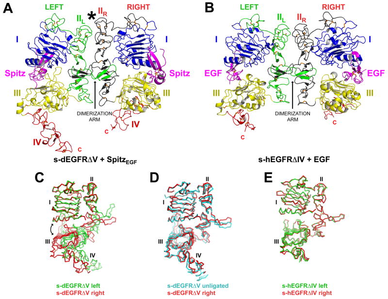
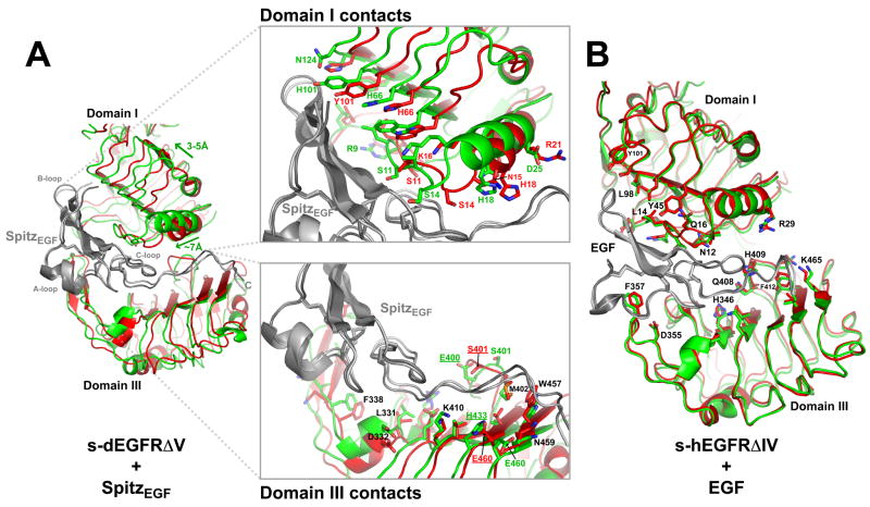
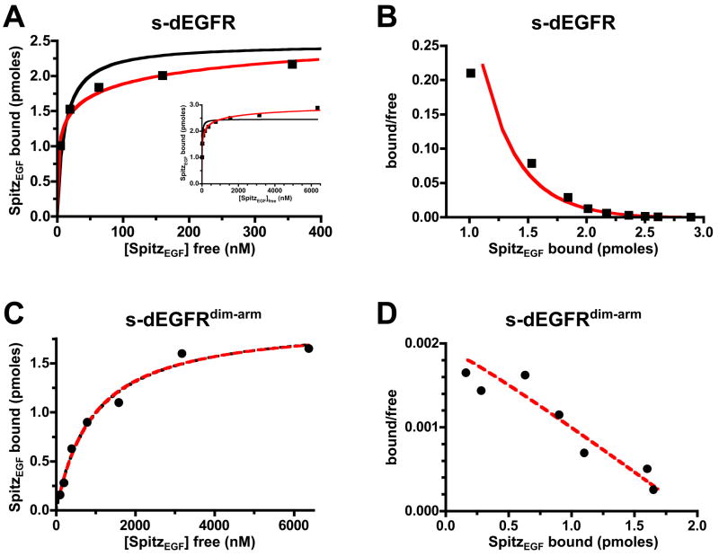
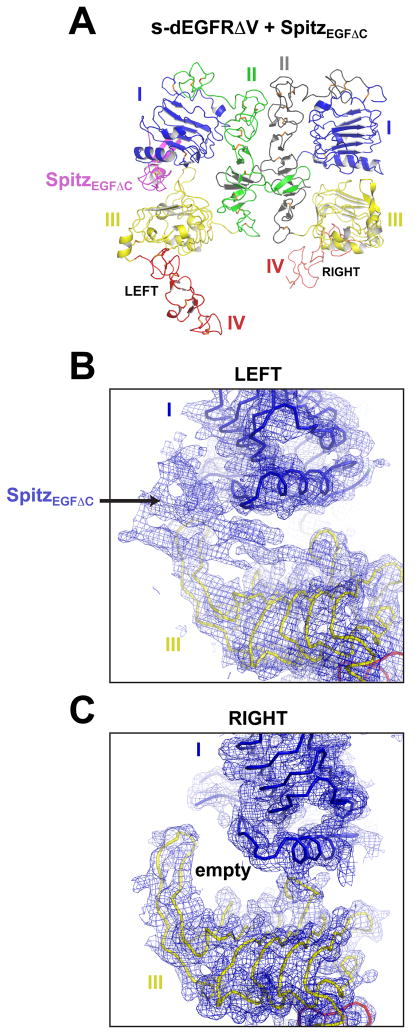
Transmembrane signaling by the epidermal growth factor receptor (EGFR) involves ligand-induced dimerization and allosteric regulation of the intracellular tyrosine kinase domain. Crystallographic studies have shown how ligand binding induces dimerization of the EGFR extracellular region but cannot explain the "high-affinity" and "low-affinity" classes of cell-surface EGF-binding sites inferred from curved Scatchard plots. From a series of crystal structures of the Drosophila EGFR extracellular region, we show here how Scatchard plot curvature arises from negatively cooperative ligand binding. The first ligand-binding event induces formation of an asymmetric dimer with only one bound ligand. The unoccupied site in this dimer is structurally restrained, leading to reduced affinity for binding of the second ligand, and thus negative cooperativity. Our results explain the cell-surface binding characteristics of EGF receptors and suggest how individual EGFR ligands might stabilize distinct dimeric species with different signaling properties.


Figures

FIGURE 1
FIGURE 1. An Asymmetric Ligand-Induced s-dEGFRΔV Dimer
(A) The (SpitzEGF)2 • (s-dEGFRΔV)2 dimer is asymmetric. Domains I, III, and IV are blue, yellow and red respectively. Domain II is green in the left-hand molecule (IIL) and dark grey in the right-hand molecule (IIR). Bound ligand is magenta. The domain II dimerization arm is labeled. An asterisk marks the amino-terminal part of domain II where asymmetry is most evident. (B) Structure (PDB code 1IVO) of the symmetric EGF-induced dimer of the human EGFR extracellular region (s-hEGFR) lacking domain IV (Ogiso et al., 2002), colored as in (A). (C) Overlay of the left (green) and right (red) molecules from the s-dEGFRΔV dimer, using domain I as reference. A double-headed curved arrow illustrates ‘wedging’ apart of domains I and III in the green molecule compared with the red molecule, breaking direct domain I/III interactions detailed in Figure S1, and altering the domain II conformation so that the dimerization arm is substantially reoriented. (D) Overlay of the right-hand molecule from the asymmetric s-dEGFRΔV dimer (red) on unligated s-dEGFRΔV (cyan) from PDB code 3I2T (Alvarado et al., 2009), using domain I as reference. (E) Overlay of the two receptor molecules in the human (EGF)2 • (s-hEGFRΔIV)2 dimer. See Table S1 for crystallographic statistics.
FIGURE 2
FIGURE 2. The Ligand-Binding Sites in the (SpitzEGF)2• (s-dEGFRΔV)2 Dimer are Inequivalent
(A) Overlay of the two ligands (grey) in the (SpitzEGF)2 • (s-dEGFRΔV)2 dimer, illustrating differences in their binding sites (see also Figure S2). The green structure corresponds to the left-hand molecule in Figure 1A, and the red structure to the right-hand molecule. Green arrows denote the ~3–5 Å shift of the green domain I towards the top left of the figure and the ~7 Å translation of the N-terminal helix described in the text. A-, B-, and C-loops of the bound ligand are labeled. The upper insert details s-dEGFRΔV side-chains that interact with SpitzEGF, highlighting significant changes. The lower insert gives a similar view of domain III interactions which are only modestly changed. Residues underlined (E400, S401, H433 and E460) are mentioned in the text. (B) Analogous overlay of the two bound ligands in the human (EGF)2 • (s-hEGFRΔIV)2 dimer from Figure 1B (Ogiso et al., 2002), illustrating similarity of the two binding sites. Most side chains that contact bound ligand overlay very well in this superimposition.
FIGURE 3
FIGURE 3. SpitzEGF Binding to s-dEGFR Yields Curved Scatchard Plots
(A) Experimental data for binding of fluorescently-labeled SpitzEGF to biotinylated s-dEGFR are well fit by the Hill equation with a Hill coefficient (nH) of 0.31 (red curve), but not a simple hyperbola (black). The inset shows saturation at >6 μM SpitzEGF. Data are representative of over six independent experiments. (B) Scatchard transformation of binding data shown in (A). The characteristic concave-up curvature is fit well by the Hill equation (nH = 0.31) – suggesting negative cooperativity. (C) Data for fluorescent SpitzEGF binding to a dimerization-defective s-dEGFR variant (s-dEGFRdim-arm) are well fit by a simple hyperbolic binding curve (black) or by the Hill equation with Hill coefficient of 1.02, suggesting no cooperativity. Data are representative of over six independent experiments. (D) Scatchard transformation of data shown in (C) yields a straight line, arguing that s-dEGFR dimerization is required for negative cooperativity.
FIGURE 4
FIGURE 4. Half-of-the-Sites Reactivity in s-dEGFRΔV
(A) Crystal structure of an s-dEGFRΔV dimer bound to SpitzEGFΔC. Ligand is bound to the left-hand molecule in which domains I and III are ‘wedged’ apart, but not the right-hand receptor molecule, which structurally resembles unligated s-dEGFRΔV. SpitzEGFΔC lacks six amino acids from its C-terminus, and binds s-dEGFRΔV with apparent KD = 4.37 ± 0.26 μM, 12-fold weaker than the value of 368 ± 23 nM measured for SpitzEGF (Figure S3). (B) Electron density is shown from a 2Fo-Fc map (blue) contoured at 1.0σ, calculated with model phases from the receptor molecules alone. In the region corresponding to the left-hand binding site in (A), clear density for bound ligand is seen. Cα traces for domains I and III are shown in blue and yellow respectively in the density, and the small part of domain IV seen is red. (C) By contrast, the 2Fo-Fc map suggests no density for bound ligand in the region corresponding to the right-hand binding site in (A). This binding site appears to be vacant in crystals of a SpitzEGFΔC • (s-dEGFRΔV)2 dimer.
FIGURE 5
FIGURE 5. Ligand Binding Promotes an Extensive Asymmetric Dimerization Interface
(A) Crystallographic dimer of unligated s-dEGFRΔV reported previously (Alvarado et al., 2009), shown surface-rendered with individual domains colored as in Figure 1A. (B) Close-up of the unligated s-dEGFRΔV dimer in the domain II region. Disulfide-bonded modules m2 to m8 are labeled, as are selected residues that interact across the ligated dimer interface in (D). An arrow marks the location between modules m4 and m5 of the ligand-induced kink (of ~12 ) that allows the amino-terminal region of the left-hand domain II to ‘collapse’ intoits right-hand counterpart in (D). (C) Surface rendered asymmetric (SpitzEGF)2 • (s-dEGFRΔV)2 dimer, with individual domains and ligand colored as in (A). (D) Domain II region close-up of the (SpitzEGF)2 • (s-dEGFRΔV)2 dimer. Disulfide-bonded modules m2, m3 and m4 from the left-hand molecule (green) have ‘collapsed’ onto their counterparts in the right-hand molecule (grey), burying 1,160 Å2 in an intimate domain II interface. Dimerization arm-mediated contacts are largely unaltered.
FIGURE 6
FIGURE 6. Model for Negatively Cooperative Ligand Binding to s-dEGFR
(A–C) Structures and cartoons describe a model for negatively cooperative ligand binding to s-dEGFRΔV. Domains (and ligand) are colored as in Figure 1. (A) Binding of a single ligand to either ‘pre-formed’ s-dEGFRΔV dimers (which have two identical binding sites) or s-dEGFRΔV monomers yields the singly ligated dimer shown in (B). (B) Singly ligated s-dEGFRΔV dimers are asymmetric. Binding of SpitzEGFΔC to the left-hand molecule wedges apart domains I and III (blue and yellow), and thus ‘bends’ domain II such thatit collapses against its counterpart in the neighboring right-hand molecule, as in Figure 5D. (C) A second SpitzEGF binds to the singly-ligated dimer, and occupies the binding site in the right-hand molecule with no change in s-dEGFR conformation. The intimate dimer interface in (B) restrains domain II in the right hand molecule, so that domains I and III cannot readily be wedged apart. Thus, the binding event that occurs in going from (B) to (C) involves a compromised set of ligand/receptor interactions as described in Figure 2A, reducing binding affinity (and retaining asymmetry in the doubly-ligated dimer). (D) The dimer of human sEGFRΔIV formed upon EGF binding is symmetric (Ogiso et al., 2002), with both ligands bound in the same manner (Figure 2B). A symmetric dimer of this sort would form following ligand binding to the dimer in (B) if ligand/receptor contacts were maximized at the expense of contacts in the dimerization interface.

References

    1. Adams TE, Koziolek EJ, Hoyne PH, Bentley JD, Lu L, Lovrecz G, Ward CW, Lee FT, Scott AM, Nash AD, et al. A truncated soluble epidermal growth factor receptor-Fc fusion ligand trap displays anti-tumour activity in vivo. Growth Factors. 2009;27:141–154. - PubMed
    1. Alvarado D, Klein DE, Lemmon MA. ErbB2 resembles an autoinhibited invertebrate epidermal growth factor receptor. Nature. 2009;461:287–291. - PMC - PubMed
    1. Bass J, Kurose T, Pashmforoush M, Steiner DF. Fusion of insulin receptor ectodomains to immunoglobulin constant domains reproduces high-affinity insulin binding in vitro. J Biol Chem. 1996;271:19367–19375. - PubMed
    1. Bellot F, Moolenaar W, Kris R, Mirakhur B, Verlaan I, Ullrich A, Schlessinger J, Felder S. High-affinity epidermal growth factor binding is specifically reduced by a monoclonal antibody, and appears necessary for early responses. J Cell Biol. 1990;110:491–502. - PMC - PubMed
    1. Bouyain S, Longo PA, Li S, Ferguson KM, Leahy DJ. The extracellular region of ErbB4 adopts a tethered conformation in the absence of ligand. Proc Natl Acad Sci U S A. 2005;102:15024–15029. - PMC - PubMed

Publication types