. 2010 Aug 20;142(4):637-46.
doi: 10.1016/j.cell.2010.07.014.

Activation of specific apoptotic caspases with an engineered small-molecule-activated protease

Affiliations

Activation of specific apoptotic caspases with an engineered small-molecule-activated protease

Daniel C Gray et al. Cell. .

Abstract

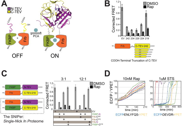
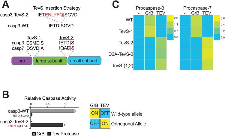
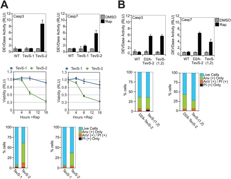
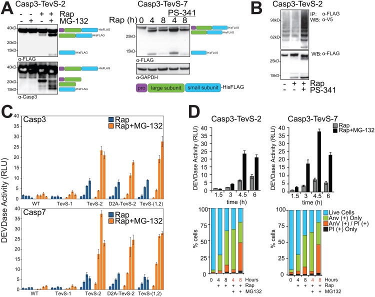
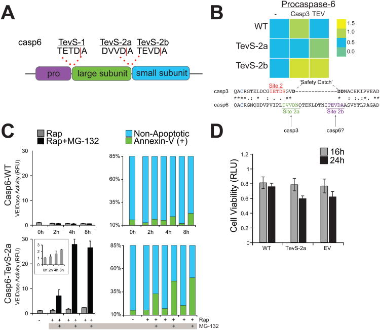
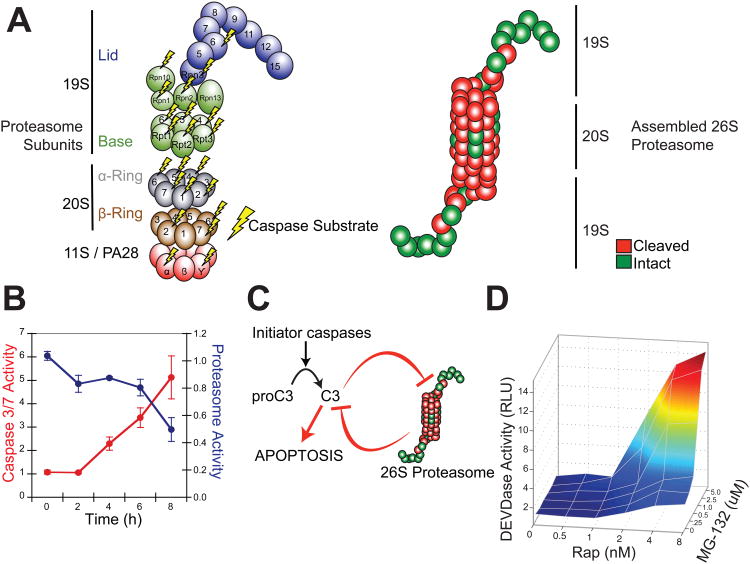
Apoptosis is a conserved cellular pathway that results in the activation of cysteine-aspartyl proteases, or caspases. To dissect the nonredundant roles of the executioner caspase-3, -6, and -7 in orchestrating apoptosis, we have developed an orthogonal protease to selectively activate each isoform in human cells. Our approach uses a split-tobacco etch virus (TEV) protease under small-molecule control, which we call the SNIPer, with caspase alleles containing genetically encoded TEV cleavage sites. These studies reveal that all three caspases are transiently activated but only activation of caspase-3 or -7 is sufficient to induce apoptosis. Proteomic analysis shown here and from others reveals that 20 of the 33 subunits of the 26S proteasome can be cut by caspases, and we demonstrate synergy between proteasome inhibition and dose-dependent caspase activation. We propose a model of proteolytic reciprocal negative regulation with mechanistic implications for the combined clinical use of proteasome inhibitors and proapoptotic drugs.


Figures

Figure 1
Figure 1. Engineering the SNIPer for Conditional Proteolysis
(A) Generalized scheme for a ligand-inducible orthogonal protease based on the protein complementation system. (B) Plasmids with deletions at the C-terminus of Frb-C-TEV or empty vector (EV) were co-transfected with FKBP-N-TEV and ECFP-TevS-YPET in 293T cells. 10nM Rap was added for 12h and FRET measurements were recorded using a fluorescent microplate reader. Data are presented as corrected averages of FRET/ECFP from an experiment performed in quadruplicate (Error Bars±SD). (C) N-TEV and C-TEV-219 were fused to either FKBP or Frb and assayed by FRET in quadruplicate as above. Transfections were performed with 3× or 12× molar excess of Tev constructs to reporter plasmid (Error Bars±SD). (D) The kinetics and cell-to-cell heterogeneity of SNIPer-mediated protease activity were compared to endogenous caspase activation by live-cell fluorescent imaging. HEK293 cells were transfected with the SNIPer and ECFP-TevS-YPET or ECFP-DEVDR-YPET, a reporter for caspase-3/-7 activity in cells, and treated with 10nM Rap or 1uM staurosporine. Live-cell FRET measurements were recorded every 15 minutes. See also Figure S1.
Figure 2
Figure 2. Design and In Vitro Characterization of Orthogonal Procaspase-3 and -7 Alleles
(A) We employed the indicated TevS insertion strategy to convert caspase sites into SNIPer-cleavage sites as shown for caspase-3. The sequences (P4-P1′ with a red bar to denote the scissile bond) and location of site-1 and site-2 are shown in the context of executioner caspase domain structure below. (B) In vitro activation of wild-type procaspase-3 (Casp3-WT) or Casp3-TevS was assayed by expressing each allele in E. coli, treating with either rat Granzyme-B (20nM) or TEV Protease(1.1uM) for 1h, and measuring caspase activity with 10uM Ac-DEVD-AFC. Data are presented as the average rate of substrate cleavage (RFU s-1) from an experiment performed in triplicate (Error Bars±SD). (C) Caspase activity of the procaspase-3 and -7 matrix of orthogonal alleles was performed as above and is presented as a heat map. Abbreviations: WT, wild-type; TevS-1, TevS insertion at site-1; TevS-2, TevS insertion at site-2; D2A-TevS-2, TevS insertion at site-2 with a non-cleavable site-1; and TevS-(1,2), TevS insertions at site-1 and site-2. See also Figure S2.
Figure 3
Figure 3. Phenotypic Analysis of SNIPer-Mediated Activation of Procaspase-3 and -7 in Cells
(A) HEK293 cells stably expressing the SNIPer and indicated procaspase-3/-7 alleles were treated with DMSO or 10nM Rap over the course of 16 hr. The indicated cells lines were assayed for caspase activity using the Caspase-Glo-3/-7 assay at 6 hr (top row), for total cellular viability using the Cell-Titer-Glo assay the indicated times (line graph, bottom row), and for phosphatidylserine (PS) exposure/membrane integrity by staining with GFP-Annexin-V and propidium iodide (PI) and imaging by fluorescent microscopy at 8 hr. Each data point is presented as an average of triplicate experiments (Error Bars±SD). (B) HEK293 stably expressing procaspase-3/-7 D2A-TevS and TevS-(1,2) alleles were analyzed for caspase activity and for GFP-Annexin-V/PI staining exactly as above. See also Figure S3.
Figure 4
Figure 4. Caspase-3 and -7 Regulation by the Proteasome
(A) The Casp3-TevS-2 line was treated with DMSO or 10nM Rap ±5uM MG-132 for 6 hr and cell lysates were blotted with anti-FLAG M2 (left panel, top) or anti-caspase-3 (left panel, bottom) antibodies. The expected processing intermediates detected by each antibody are shown to the right of the immunoblots. The Casp7-TevS-2 line was treated with 10nM Rap for 4 or 8 hr ±500nM PS-341 and immunoblotted with anti-FLAG M2 (right panel) and anti-GAPDH. (B) The Casp3-TevS-2 line was transfected with pcDNA3.1-V5-Ubiquitin for 24 hr and treated with DMSO, 10nM Rap or 10nM Rap + 500nM PS-341 for 4 hr. Cell lysates were immunoprecipitated with anti-FLAG M2 agarose and immunoblotted for covalent ubiquitin with an anti-V5 antibody or with anti-flag to assess caspase-3 recovery. (C) The Casp3-TevS-2 and Casp7-TevS-2 lines were treated with 10nM Rap (blue bars) or 10nM Rap and 5uM MG-132 (orange bars). Caspase activity was measured every 1.5 hr in triplicate (Error Bars±SD). (D) Casp3-TevS-2 and Casp7-TevS-7 cell lines were treated with 10nM Rap ±5uM MG-132 and assayed for caspase activity as above (top) or by staining with GFP-Annexin-V and PI and imaging with fluorescent microscopy (bottom) for indicated time courses in triplicate (Error Bars±SD). See also Figure S4.
Figure 5
Figure 5. Proteasome Inhibition Converts Procaspase-6 to an Apoptotic Executioner Caspase Isoform
(A) Sites for which the TevS insertion was applied for caspase-6. (B)In vitro caspase activity of the procaspase-6 matrix of orthogonal alleles is presented as a heat map (top). Caspase activity was measured by expressing each allele in E. coli, treating with active caspase-3 (2nM) or Tev Protease(1.1uM) for 1h with 30uM Ac-VEID-AFC in triplicate, correcting for caspase-3 VEIDase activity where appropriate. Abbreviations: WT, wild-type; TevS-2a, TevS insertion at site-2a with P1′ Ala; TevS-2b, TevS insertion at site-2b. Alignment of the protein sequences of procaspase-3 (CASP3_HUMAN) and procaspase-6 (CASP6_HUMAN) indicates an extra loop region in caspase-6 that harbors site-2a and -2b. (C) The Casp6-WT (top) line was treated with 10nM Rap ±5uM MG-132 over a course of 8h and assayed for caspase-6 activity with 50uM VEID-R110 (left) or by staining with GFP-Annexin-V imaging with fluorescent microscopy. Casp6-TevS-2 (bottom) was treated identically. The inset is the same assay in the range of 0-3 RFU. Data are presented as the average from an experiment performed in quadruplicate (Error Bars±SD). (D) The Casp3-TevS-2 line was treated with 10nM Rap for 16 or 24 hr and cell viability was measured with Cell Titer-Glo (Promega, WI). See also Figure S5.
Figure 6
Figure 6. Reciprocal Negative Regulation between the 26S Proteasome and Executioner Caspases
(A) Novel and published proteasome subunits identified as caspase substrates are mapped to an inventory of 20S core, 19S lid/base and 11S activator subunits (left). Cleaved subunits (red) are also mapped onto a model of the fully assembled, multimeric 26S proteasome (right). (B) The Casp3-TevS-2 line was treated with 10nM Rap over a course of 8 hr and assayed for caspase activity with Caspase-Glo-3/-7 and protease activity with Proteasome-Glo (Chymotrypsin-Like). Data are presented as the average from an experiment performed in triplicate (Error Bars±SD; note different scales). (C) A model for reciprocal negative regulation between activated executioner caspases and the 26S proteasome-ubiquitin system predicts synergy between caspase activation and proteasome inhibition. (D) The Casp3-TevS-2 line was co-treated with the indicated concentrations of Rap (0-8nM) and MG-132 (0-5uM) for 4 hr and caspase activation was measured with Caspase-Glo-3/-7 in triplicate. See also Table S1.

Comment in

  • SNIPer pulls the trigger.
    Doerr A. Doerr A. Nat Methods. 2010 Oct;7(10):786. doi: 10.1038/nmeth1010-786. Nat Methods. 2010. PMID: 20936776 No abstract available.

References

    1. Adrain C, Creagh EM, Cullen SP, Martin SJ. Caspase-dependent inactivation of proteasome function during programmed cell death in Drosophila and man. J Biol Chem. 2004;279:36923–36930. - PubMed
    1. Albeck JG, Burke JM, Aldridge BB, Zhang M, Lauffenburger DA, Sorger PK. Quantitative analysis of pathways controlling extrinsic apoptosis in single cells. Mol Cell. 2008a;30:11–25. - PMC - PubMed
    1. Albeck JG, Burke JM, Spencer SL, Lauffenburger DA, Sorger PK. Modeling a snap-action, variable-delay switch controlling extrinsic cell death. Plos Biol. 2008b;6:2831–2852. - PMC - PubMed
    1. Bedford L, Hay D, Devoy A, Paine S, Powe DG, Seth R, Gray T, Topham I, Fone K, Rezvani N, et al. Depletion of 26S proteasomes in mouse brain neurons causes neurodegeneration and Lewy-like inclusions resembling human pale bodies. J Neurosci. 2008;28:8189–8198. - PMC - PubMed
    1. Casciola-Rosen L, Garcia-Calvo M, Bull H, Becker J, Hines T, Thornberry N, Rosen A. Mouse and Human Granzyme B Have Distinct Tetrapeptide Specificities and Abilities to Recruit the Bid Pathway. Journal of Biological Chemistry. 2007;282:4545. - PubMed

Publication types