. 2010 Aug 24;56(9):721-34.
doi: 10.1016/j.jacc.2010.03.066.

Guided cardiopoiesis enhances therapeutic benefit of bone marrow human mesenchymal stem cells in chronic myocardial infarction

Affiliations

Guided cardiopoiesis enhances therapeutic benefit of bone marrow human mesenchymal stem cells in chronic myocardial infarction

Atta Behfar et al. J Am Coll Cardiol. .

Abstract

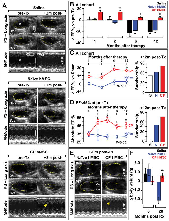
Objectives: The goal of this study was to guide bone marrow-derived human mesenchymal stem cells (hMSCs) into a cardiac progenitor phenotype and assess therapeutic benefit in chronic myocardial infarction.

Background: Adult stem cells, delivered in their naïve state, demonstrate a limited benefit in patients with ischemic heart disease. Pre-emptive lineage pre-specification may optimize therapeutic outcome.

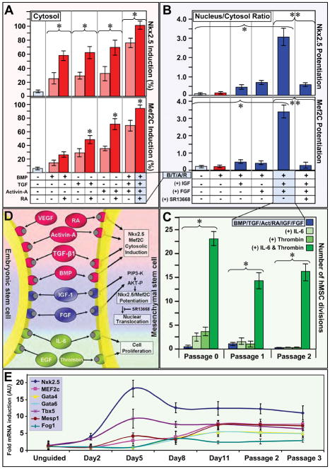
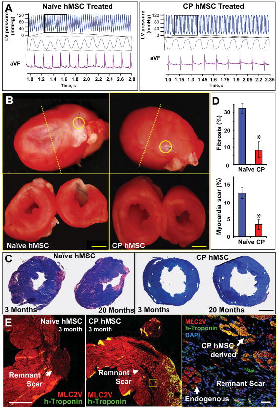
Methods: hMSC were harvested from a coronary artery disease patient cohort. A recombinant cocktail consisting of transforming growth factor-beta(1), bone morphogenetic protein-4, activin A, retinoic acid, insulin-like growth factor-1, fibroblast growth factor-2, alpha-thrombin, and interleukin-6 was formulated to engage hMSC into cardiopoiesis. Derived hMSC were injected into the myocardium of a nude infarcted murine model and followed over 1 year for functional and structural end points.

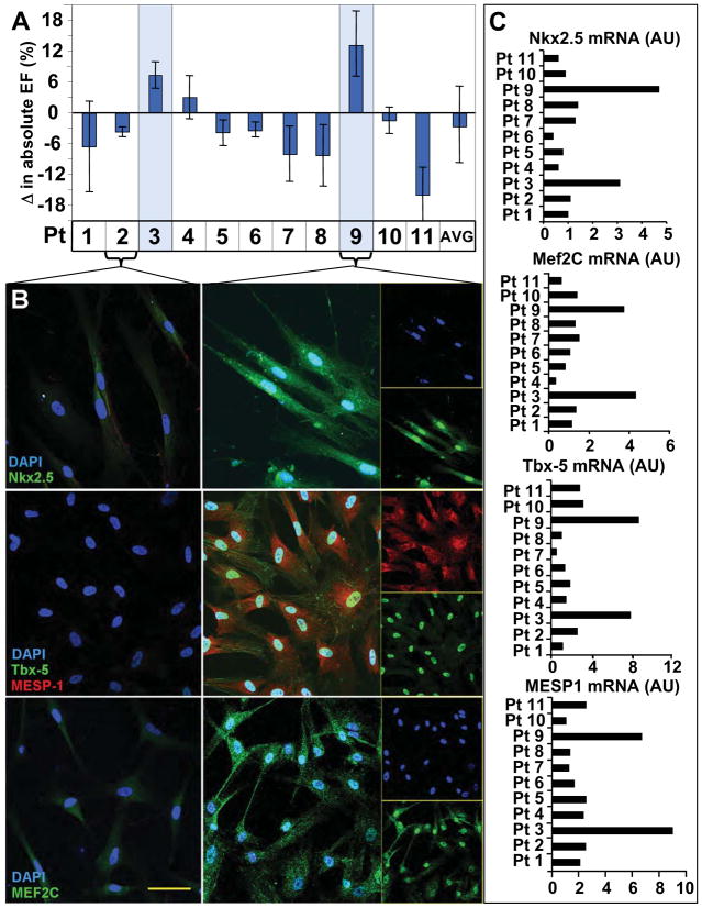
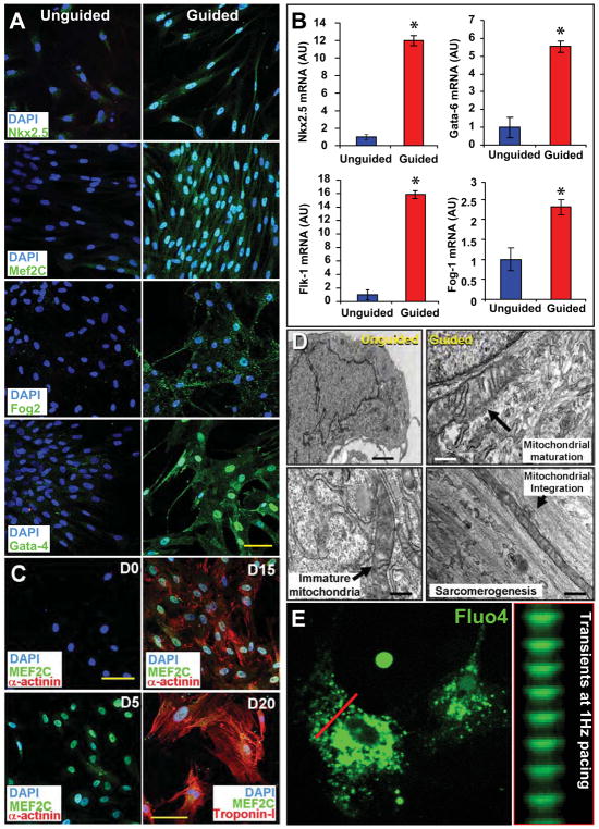
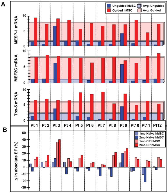
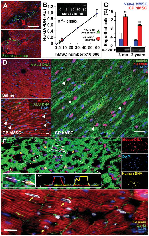
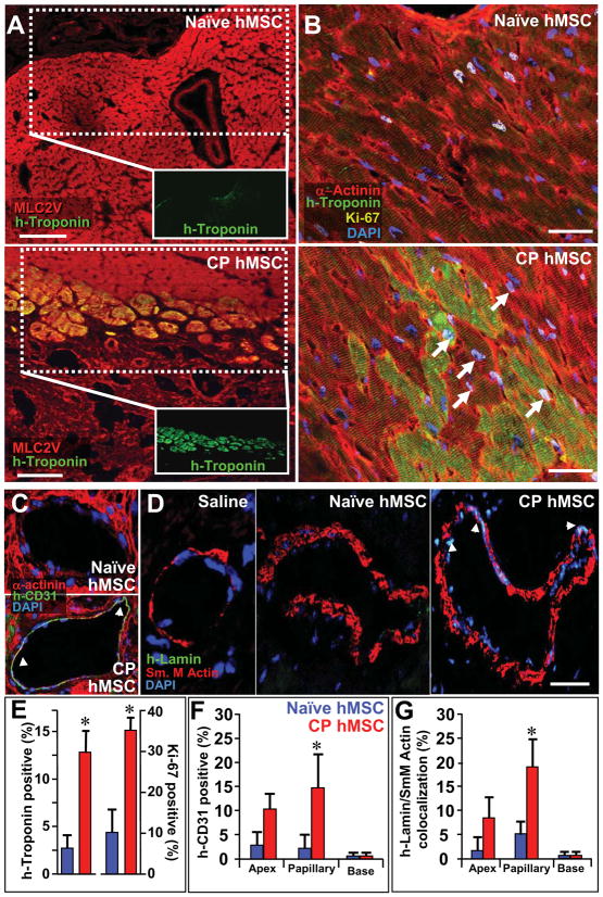
Results: Although the majority of patient-derived hMSC in their native state demonstrated limited effect on ejection fraction, stem cells from rare individuals harbored a spontaneous capacity to improve contractile performance. This reparative cytotype was characterized by high expression of homeobox transcription factor Nkx-2.5, T-box transcription factor TBX5, helix-loop-helix transcription factor MESP1, and myocyte enhancer factor MEF2C, markers of cardiopoiesis. Recombinant cardiogenic cocktail guidance secured the cardiopoietic phenotype across the patient cohort. Compared with unguided counterparts, cardiopoietic hMSC delivered into infarcted myocardium achieved superior functional and structural benefit without adverse side effects. Engraftment into murine hearts was associated with increased human-specific nuclear, sarcomeric, and gap junction content along with induction of myocardial cell cycle activity.

Conclusions: Guided cardiopoiesis thus enhances the therapeutic benefit of bone marrow-derived hMSC in chronic ischemic cardiomyopathy.


Conflict of interest statement

Conflict of Interest:

A.B., J.B., and A.T. serve on the research advisory board of Cardio3 BioSciences.

Figures

Figure 1
Figure 1. Repair efficacy in hMSC correlates with cardiac transcription factor expression
A, Patient hMSC demonstrated variable repair after injection into an infarction model. Only rare stem cells (patients #3 and #9 highlighted) demonstrated benefit. B, Low expression of cardiac transcription factors in non-reparative hMSC (patient #2 illustrated). Robust Nkx2.5 (83±5% and 86±2% of hMSC in patients #3 and #9), MEF2C, Tbx-5, and MESP-1 expression in hMSC with reparative capacity (patient #9 shown, with single channels provided in inset). Bar, 20 μm. C, Reparative pattern validated with upregulated Nkx2.5, Mef2c, Tbx-5 and MESP1 mRNA in reparative versus non-reparative hMSC.
Figure 2
Figure 2. Derivation of cardiogenic cocktail that induces and potentiates cardiac transcription factor expression, and maintains cell cycle activity
A, BMP-4, TGFβ1, Activin-A induced cytosolic expression of Nkx2.5 and Mef2C (light red columns). Addition of retinoic acid (RA) boosted cytosolic induction (red columns). B, BMP-4, TGFβ1, Activin-A and RA (B/T/A/R, red) potentiated nuclear translocation of Nkx2.5 and Mef2C when combined with IGF-1 and FGF-2. AKT inhibitor (SR13668) inhibited nuclear translocation. C, Low hMSC cell cycle activity following BMP-4, TGFβ1, Activin-A, RA, IGF-1, and FGF-2 treatment (blue) rescued by IL-6 and human α-thrombin. Brackets indicate pared analysis. Star and double star, p<0.05. D, Cardiogenic cocktail, applied as a combined regimen, induced and potentiated nuclear translocation of Nkx2.5 and Mef2C while maintaining cell cycle activity. E, Nkx2.5, Tbx5, MEF2C, GATA4, GATA6, FOG1 and MESP1 hMSC mRNA in response to cocktail treatment. Panels A-C and E evaluate non-reparative patient hMSC.
Figure 3
Figure 3. Cardiogenic cocktail guides cardiogenesis to yield functional cardiomyocytes
A, Nuclear translocation of Nkx2.5, Mef2C, Fog-2, and Gata-4 in cocktail-guided hMSC. B, Guidance of hMSC, from 12 coronary artery disease patients, increased mRNA expression of Nkx2.5, Flk-1, Gata-6, and Fog-1. Star, p<0.01. C, Cocktail-based cardiogenic conversion of naïve hMSC (D0) resulted in Mef2C activation at day 5 (D5), and sarcomerogenesis (α-Actinin) achieved by day 15 (D15) to 20 (D20, Troponin I expression). Bar, 20 μm. D, Compared to naïve hMSC with abundant nuclei (Left), transmission electron microscopy of cocktail-guided hMSC at day 15 revealed mitochondrial maturation and integration with organizing myofibrils. Bar, 1 μm (upper left); 250 nm (other panels). E, Calcium transients elicited in response to 1-Hz electrical stimulation. See also Supplemental Movie depicting calcium transients in Fluo4AM-loaded and paced cardiopoietic hMSC at 20 days following 1% platelet lysate differentiation.
Figure 4
Figure 4. Cardiopoietic hMSC phenotype associated with ejection fraction improvement
A, Induction of cardiac transcription factors Tbx-5, MEF2c, and MESP-1 mRNA documented for each patient. B, Echocardiography demonstrated ejection fraction improvement in chronically infarcted mice treated with cardiopoietic (CP) over patient-matched naïve hMSC at 1–2 months. Experiment in B distinct from Figure 1A.
Figure 5
Figure 5. Cardiopoietic (CP) hMSC provide benefit following transplantation
A, Echocardiography of infarcted hearts 4 weeks after coronary ligation, prior to therapy (pre-Tx) revealed an akinetic anterior wall. Saline and naïve hMSC treated hearts maintained akinesis in the anterior wall, in contrast to re-animation in CP treated (+2m post-Tx, arrowhead). See Supplemental Movies depicting anterior wall reanimation in the long and short axis on echocardiography from cardiopoietic but not naïve hMSC-treated cohorts at 1-year post-therapy. B, Cardiopoietic transplantation (CP; n=14) was associated with ejection fraction improvement acutely and limited progression towards failure chronically, not observed in saline (S; n=10) or naïve groups (N; n=17). C, Sustained benefit of CP hMSC therapy over 1-year follow-up, compared to naïve hMSC treatment, was associated with survival benefit (right). Ejection fraction expressed relative to sham (dotted line). D, Subgroup analysis of mice with heart failure (pre-Tx ejection fraction <45%), CP hMSC had superior benefit versus naïve, with maintained survival (right). Saline treated mice in this subgroup did not survive beyond 2 months. E-F, CP hMSC treatment prevented heart failure and blunted weight loss, compared to naïve (n=5) at 20 months. In E, LV, left ventricle; Ao, aorta; LA, left atrium. Dias, diastole; Sys, systole. In E, arrowhead indicates reanimated anterior wall. Error bars represent SEM. Star, p<0.01 in B, C and D; p<0.05 in F.
Figure 6
Figure 6. Echocardiographic improvement validated with pressure catheter and histopathological evaluation
A, Catheterization revealed no afterload discrepancy between cohorts, yet elevated end-diastolic pressures were noted in naïve versus cardiopoietic (CP) hMSC groups. B, Pathological evaluation demonstrated diminished scar downstream of left anterior descending (LAD) artery ligation (yellow circle) with re-muscularization and diminished remodeling in CP (right) in contrast to naïve (left) treated hearts at 3-months (yellow circle). C, Diffuse fibrosis and larger scar area at 3 and 20 months in naïve compared to CP hMSC treated hearts. Bar, 2 mm for B and C. D, Scar size and total myocardial fibrosis (n=10 per condition). Star, p<0.01. E, Immunohistological evaluation of scar reveals limited contribution of human-troponin cells in naïve (left) in contrast to cardiopoietic treated hearts (middle, right). Bar, 500 μm - left and 20 μm - right.
Figure 7
Figure 7. Cardiopoietic (CP) hMSC demonstrate long-term integration
A, Fluorochrome loaded CP hMSC detected within murine myocardium 48 h after delivery. B, mRNA standard curve for human-specific GAPDH with escalating doses of hMSC within murine myocardium. Quantified hMSC engraftment (3-month, red circle; 2-years, green triangle). C, Higher CP hMSC engraftment compared to naïve. Star, p<0.01. D, Compared to saline (left), CP hMSC within treated hearts confirmed by human h-ALU DNA (green, middle) and immuno-probing for human-specific lamin (red, right). E, Concurrent human (yellow) and mouse (red) DNA probing demonstrated negligible fusion through species-specific nuclear staining on confocal histogram and single channel analysis (insets). Bar, 20 μm for A, D, E. F, Human lamin (yellow, arrows) expressing myocytes demonstrated connexin-43 (green) expression. Bar, 10 μm.
Figure 8
Figure 8. Cardiopoietic (CP) hMSC demonstrate cardiogenic and vasculogenic potential within host myocardium
A, Human-specific troponin-I (green) in the anterior wall of naive- versus CP-treated hearts, respectively, co-localized with ventricular myosin light chain (MLC2v, red). Bar, 100 μm. B, Human troponin-I staining of naïve versus CP hMSC treated hearts, counterstained with α-Actinin (red), demonstrated engraftment of human cells. Cell cycle activation, documented by Ki-67 expression (yellow, arrows), noted in human troponin positive and endogenous cardiomyocytes. C, Confocal evaluation of collateral vessels from CP hMSC treated hearts demonstrated human-specific CD-31 (PECAM-1) staining. D, Human lamin staining (arrows) co-localized with nuclei of smooth muscle in vessels from CP hMSC treated but not saline or naïve treated hearts. Bar, 20 μm for B-D. E, Percent of human troponin and Ki-67 positive cells in myocardial wall, upregulated in CP versus naïve hMSC treated hearts (n=10). F-G, Quantification of human CD-31 and co-localization between human-lamin and smooth muscle actin reveals significant contribution at the level of the papillary muscle from CP hMSC compared to naïve (n=10 in F and G). Star, p<0.05.

Comment in

  • Boot camp for mesenchymal stem cells.
    Marbán E, Malliaras K. Marbán E, et al. J Am Coll Cardiol. 2010 Aug 24;56(9):735-7. doi: 10.1016/j.jacc.2010.02.064. J Am Coll Cardiol. 2010. PMID: 20723803 Free PMC article. No abstract available.

References

    1. Dimmeler S, Zeiher AM, Schneider MD. Unchain my heart: the scientific foundations of cardiac repair. J Clin Invest. 2005;115:572–83. - PMC - PubMed
    1. Srivastava D, Ivey KN. Potential of stem-cell-based therapies for heart disease. Nature. 2006;441:1097–9. - PubMed
    1. Segers VF, Lee RT. Stem-cell therapy for cardiac disease. Nature. 2008;451:937–42. - PubMed
    1. Abdel-Latif A, Bolli R, Tleyjeh IM, et al. Adult bone marrow-derived cells for cardiac repair: a systematic review and meta-analysis. Arch Intern Med. 2007;167:989–97. - PubMed
    1. Passier R, van Laake LW, Mummery CL. Stem-cell-based therapy and lessons from the heart. Nature. 2008;453:322–9. - PubMed

Publication types

MeSH terms