. 2010 Dec 2;101(3):251-64.
doi: 10.1016/j.jphotobiol.2010.07.010. Epub 2010 Jul 30.

The malondialdehyde-derived fluorophore DHP-lysine is a potent sensitizer of UVA-induced photooxidative stress in human skin cells

Affiliations

The malondialdehyde-derived fluorophore DHP-lysine is a potent sensitizer of UVA-induced photooxidative stress in human skin cells

Sarah D Lamore et al. J Photochem Photobiol B. .

Abstract

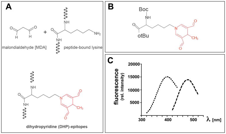
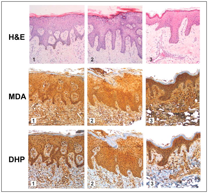
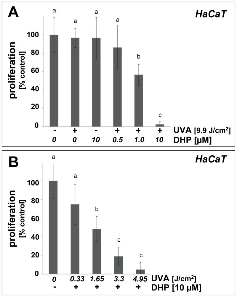
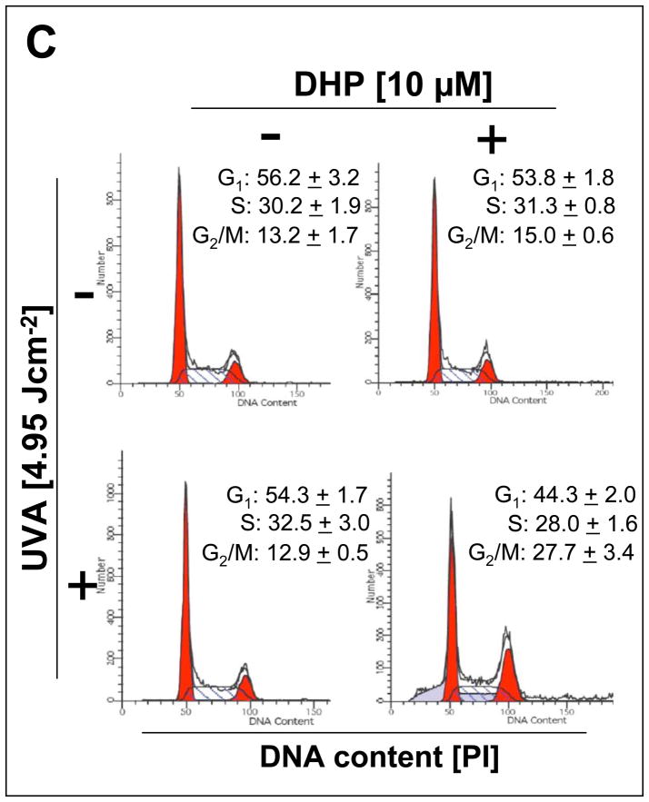
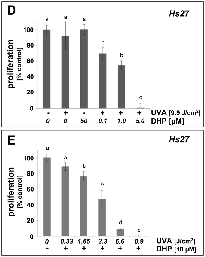
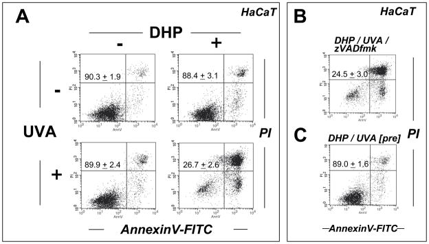
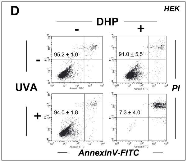
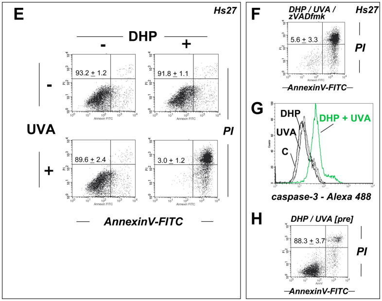
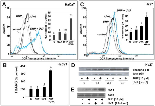
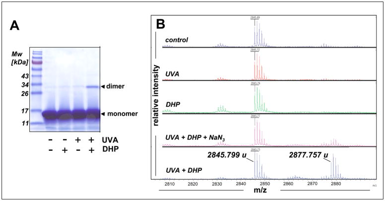
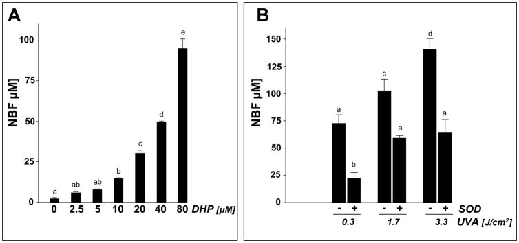
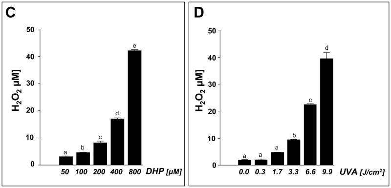
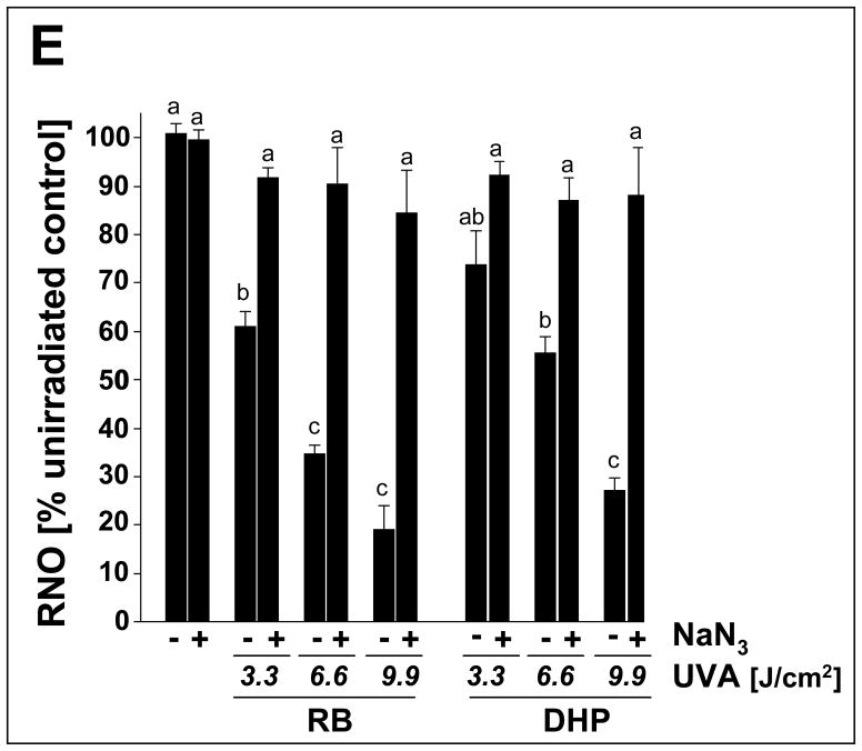
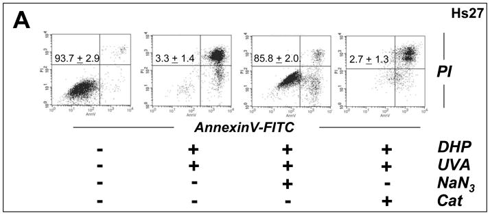
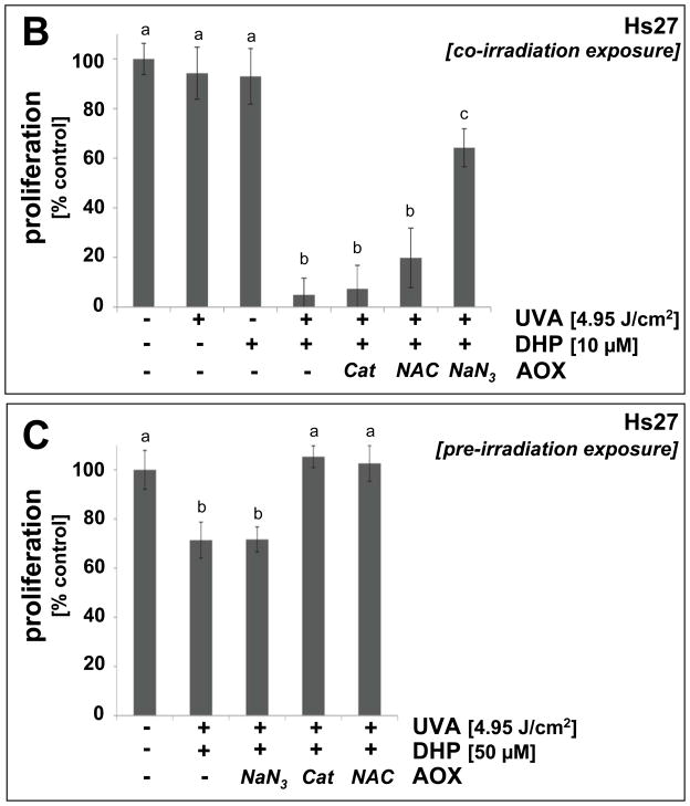
Light-driven electron and energy transfer involving non-DNA skin chromophores as endogenous photosensitizers induces oxidative stress in UVA-exposed human skin, a process relevant to photoaging and photocarcinogenesis. Malondialdehyde is an electrophilic dicarbonyl-species derived from membrane lipid peroxidation. Here, we present experimental evidence suggesting that the malondialdehyde-derived protein epitope dihydropyridine (DHP)-lysine is a potent endogenous UVA-photosensitizer of human skin cells. Immunohistochemical analysis revealed the abundant occurrence of malondialdehyde-derived and DHP-lysine epitopes in human skin. Using the chemically protected dihydropyridine-derivative (2S)-Boc-2-amino-6-(3,5-diformyl-4-methyl-4H-pyridin-1-yl)-hexanoic acid-t-butylester as a model of peptide-bound DHP-lysine, photodynamic inhibition of proliferation and induction of cell death were observed in human skin Hs27 fibroblasts as well as primary and HaCaT keratinocytes exposed to the combined action of UVA and DHP-lysine. DHP-lysine photosensitization induced intracellular oxidative stress, p38 MAPkinase activation, and upregulation of heme oxygenase-1 expression. Consistent with UVA-driven ROS formation from DHP-lysine, formation of superoxide, hydrogen peroxide, and singlet oxygen was detected in chemical assays, but little protection was achieved using SOD or catalase during cellular photosensitization. In contrast, inclusion of NaN(3) completely abolished DHP-photosensitization. Taken together, these data demonstrate photodynamic activity of DHP-lysine and support the hypothesis that malondialdehyde-derived protein-epitopes may function as endogenous sensitizers of UVA-induced oxidative stress in human skin.


Figures

Figure 1
Figure 1. The lipid peroxidation-derived fluorophore dihydropyridine (DHP)-lysine
(A) Formation of DHP-epitopes on target lysine residues occurs by spontaneous malondialdehyde (MDA)-adduction. (B) Protected DHP-lysine as a model of peptide-bound DHP-lysine. (C) Fluorescence spectrum of protected DHP-lysine [excitation spectrum (λem at 485 nm; broken line, short dashes) and emission spectrum (λex at 395 nm; broken line, long dashes)]
Figure 2
Figure 2. Immunohistochemical detection of MDA- and DHP-epitopes in healthy human skin
A commercially available healthy human skin tissue microarray (NS21-01-TMA, Cybrdi) was processed for H&E staining (top row, specimens 1–3), pan-MDA-immunohistochemistry (middle row, specimens 1–3) using a polyclonal antibody (AP050), and DHP-immunohistochemistry (bottom row, specimens 1–3) using a monoclonal antibody (1F83). In all specimens, abundant staining for MDA- and DHP-epitopes occurs throughout the epidermal and dermal layers; Staining is most abundant in the cellular layers of the epidermis. Stratum corneum does not stain positive for either epitope. Three representative specimens are depicted.
Figure 3
Figure 3. DHP-lysine as a photosensitizer of UVA-induced inhibition of skin cell proliferation
(A) Human immortalized HaCaT keratinocytes were exposed to UVA-irradiation (9.9 J/cm2) in the presence or absence of DHP-lysine (0.5–10 μM). Additionally, mock-irradiated cells were exposed to DHP (10 μM). Cells were washed with PBS, fresh DMEM was added, and cell number was determined 72 h later by cell counting. Proliferation was compared to untreated cells. (B) HaCaT keratinocytes were exposed to increasing doses of UVA-irradiation (up to 4.95 J/cm2) in the presence or absence of DHP-lysine (10 μM). Additionally, mock-irradiated cells were exposed to DHP (10 μM). After treatment, proliferation was assessed as detailed in panel A. (C) Cell cycle analysis by flow cytometric analysis of HaCaT keratinocytes stained with propidium iodide was performed 24h after photosensitization (DHP (10 μM); UVA (4.95 J/cm2). Histograms of a representative experiment are shown. The numbers summarize results (% of total gated cells; mean + SD) from three independent experiments. (D–E) Human skin fibroblasts (Hs27) were exposed to UVA-irradiation in the presence or absence of DHP-lysine, and effects of photosensitization on proliferation were then examined as detailed in panels A–B. In the bar graphs, means with common letter differ (p<0.05).
Figure 4
Figure 4. DHP-lysine as a photosensitizer of UVA-induced skin cell death
(A–C) HaCaT keratinocytes were exposed to the combined action of UVA irradiation (9.9 J/cm2) and DHP-lysine (50 μM). (A) Induction of cell death 24 h after exposure was examined by flow cytometric analysis of annexinV-PI stained cells. (B) Photosensitization was performed as in panel A in the presence of zVADfmk. (C) Unirradiated cells were exposed to DHP (50 μM) that was UVA-preirradiated (9.9 J/cm2) and cell viability was examined 24 h after exposure. The numbers summarize results (% viable cells (lower left quadrant) of total gated cells; mean + SD) from three independent experiments. (D) Primary human epidermal keratinocytes (HEK) were exposed to the combined action of UVA irradiation (9.9 J/cm2) and DHP-lysine (50 μM). Induction of cell death 24 h after exposure was examined by flow cytometric analysis of annexinV-PI stained cells. (E–H) Hs27 fibroblasts were exposed to the combined action of UVA irradiation (9.9 J/cm2) and DHP-lysine (50 μM). (E) Induction of cell death 24 h after exposure was examined by flow cytometric analysis of annexinV-PI stained cells. (F) Photosensitization was performed as in panel A in the presence of zVADfmk. (G) Photosensitization was performed as in panel A, and caspase 3 activation was analyzed by flow cytometry using an Alexa 488-conjugated antibody directed against cleaved procaspase 3. (H) Unirradiated cells were exposed to DHP (50 μM) that was UVA-preirradiated (9.9 J/cm2) and cell viability was examined 24 h after exposure. The numbers summarize results (% viable cells (lower left quadrant) of total gated cells; mean + SD) from three independent experiments.
Figure 5
Figure 5. Induction of oxidative stress in cultured human skin cells resulting from DHP-lysine photosensitization
(A) HaCaT keratinocytes were exposed to UVA-irradiation (9.9 J/cm2) in the presence or absence of DHP (50 μM) followed by loading with the intracellular redox dye DCFH-DA 1 h after irradiation. DCF-fluorescence intensity indicative of intracellular redox stress was then quantified by flow cytometric analysis. One representative histogram out of three similar repeats is shown. In the bar graph, means with common letter differ (n=3, mean + SD; p<0.05). (B) Lipid peroxidation resulting from DHP-lysine photosensitization was examined in HaCaT keratinocytes exposed as specified in (A) followed by photometric detection of TBARS. (C) Hs27 fibroblasts were exposed and analyzed as specified in (A). (D) Upregulation of cellular heme oxygenase-1 protein levels by the combined action of DHP-lysine and UVA was examined in Hs27 fibroblasts 24 h after photosensitization by Western blotting as described in Experimental Procedures. (E) Photosensitized induction of p38 MAPkinase phosphorylation by the combined action of DHP-lysine and UVA was assessed in Hs27 fibroblasts. 30 min after photosensitization cells were lysed and analyzed by Western blotting using polyclonal anti-phospho p38 and anti-total p38 antibodies as described in ‘Materials and methods’.
Figure 6
Figure 6. Induction and antioxidant modulation of protein and peptide photodamage sensitized by DHP-lysine
(A) Photosensitization of protein damage by DHP-lysine was assessed using an RNAse A photo-crosslinking assay. RNAse A (10 mg/mL PBS) was irradiated with UVA (9.9 J/cm2) in the absence or presence of DHP-lysine (50 μM) and a reaction aliquot was analyzed by 15% SDS-PAGE followed by Coomassie-staining [Migration positions of molecular weight standard (Mw), RNAse monomer, and RNAse dimer are indicated]. (B) Mass spectrometric analysis of peptide photooxidation sensitized by DHP-lysine. The peptide melittin (1 mg/mL PBS) was UVA-irradiated (9.9 J/cm2 UVA) in the presence or absence of DHP-lysine (10 μM) followed by MALDI-TOF mass spectrometric analysis. In an additional group, exposure to UVA plus DHP-lysine occurred in the presence of NaN3 (10 mM). Monoisotopic mass peaks are indicated.
Fig. 7
Fig. 7. DHP-lysine-sensitized production of ROS
Photosensitization of ROS formation upon exposure to UVA was examined over a wide concentration range of DHP-lysine (2.5 up to 800 μM). (A) DHP-lysine-sensitized NBT reduction measured by NBF generation as a function of DHP-lysine concentration (3.3 J/cm2 UVA). (B) SOD-suppressible NBF formation indicative of superoxide formation was assessed by exposing DHP-lysine (100 μM) to increasing doses of UVA in the presence or absence of SOD (15000 u/mL). Values represent the mean of three independent experiments + SD. (C) Dose response of H2O2 production as a function of DHP- lysine concentration (9.9 J/cm2 UVA). (D) Dose response of H2O2 production as a function of UVA dose (800 μM DHP-lysine). (E) Singlet oxygen formation as evidenced by the RNO bleaching assay. Loss of RNO absorbance resulting from photosensitization by Rose Bengal (RB, 1 μM) and DHP-lysine (20 μM) was examined as a function of UVA dose in the absence or presence of NaN3 (10 mM).
Fig. 8
Fig. 8. Antioxidant protection of human skin fibroblasts against DHP-lysine phototoxicity
(A) Human Hs27 fibroblasts were exposed to the combined action (‘co-irradiation exposure’) of UVA-irradiation (9.9 J/cm2) and DHP-lysine (50 μM) in the presence or absence of various antioxidants including NaN3 (10 mM) and catalase (Cat, 400 u/mL). Viability was examined 24 h after exposure by flow cytometric analysis of annexinV-FITC/PI-stained cells. (B) Cells were exposed to the combined action of UVA-irradiation (4.95 J/cm2) and DHP-lysine (10 μM) in the presence or absence of various antioxidants including NaN3 (10 mM), NAC (10 mM), and catalase (Cat, 400 u/mL). Proliferation was examined 72 h after exposure as specified in Materials and Methods. (C) DHP-lysine (10 μM) was exposed to UVA (9.9 J/cm2) (‘pre-irradiation exposure’) and then immediately added to un-irradiated cells in the presence or absence of various antioxidants (30 min exposure, followed by media change), and proliferation was examined 72 h later.

Similar articles

Cited by

References

    1. Scharffetter-Kochanek K, Wlaschek M, Brenneisen P, Schauen M, Blaudschun R, Wenk J. UV-induced reactive oxygen species in photocarcinogenesis and photoaging. Biol Chem. 1997;378:1247–1257. - PubMed
    1. Wondrak GT, Jacobson MK, Jacobson EL. Endogenous UVA-photosensitizers: mediators of skin photodamage and novel targets for skin photoprotection. Photochem Photobiol Sci. 2006;5:215–237. - PubMed
    1. Brash DE, Rudolph JA, Simon JA, Lin A, McKenna GJ, Baden HP, Halperin AJ, Ponten J. A role for sunlight in skin cancer: UV-induced p53 mutations in squamous cell carcinoma. Proc Natl Acad Sci U S A. 1991;88:10124–10128. - PMC - PubMed
    1. Gasparro FP. Sunscreens, skin photobiology, and skin cancer: the need for UVA protection and evaluation of efficacy. Environ Health Perspect. 2000;108(Suppl 1):71–78. - PMC - PubMed
    1. Halliday GM. Inflammation, gene mutation and photoimmunosuppression in response to UVR-induced oxidative damage contributes to photocarcinogenesis. Mutat Res. 2005;571:107–120. - PubMed

Publication types

MeSH terms

LinkOut - more resources