

Adaptation of the vestibulo-ocular reflex for forward-eyed foveate vision
- PMID: 20724359
- PMCID: PMC3000578
- DOI: 10.1113/jphysiol.2010.196287
Adaptation of the vestibulo-ocular reflex for forward-eyed foveate vision
Abstract
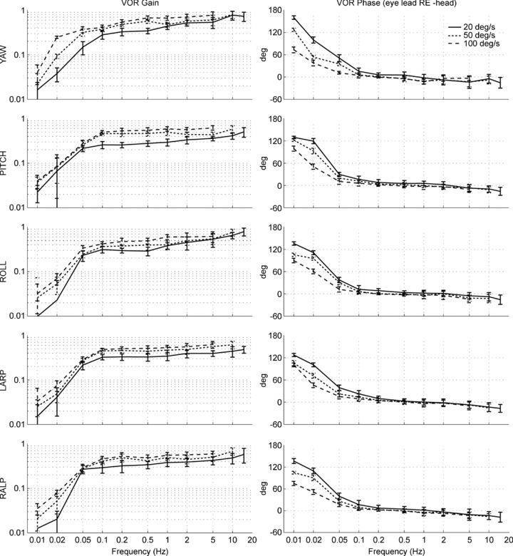
To maintain visual fixation on a distant target during head rotation, the angular vestibulo-ocular reflex (aVOR) should rotate the eyes at the same speed as the head and in exactly the opposite direction. However, in primates for which the 3-dimensional (3D) aVOR has been extensively characterised (humans and squirrel monkeys (Saimiri sciureus)), the aVOR response to roll head rotation about the naso-occipital axis is lower than that elicited by yaw and pitch, causing errors in aVOR magnitude and direction that vary with the axis of head rotation. In other words, primates keep the central part of the retinal image on the fovea (where photoreceptor density and visual acuity are greatest) but fail to keep that image from twisting about the eyes' resting optic axes. We tested the hypothesis that aVOR direction dependence is an adaptation related to primates' frontal-eyed, foveate status through comparison with the aVOR of a lateral-eyed, afoveate mammal (Chinchilla lanigera). As chinchillas' eyes are afoveate and never align with each other, we predicted that the chinchilla aVOR would be relatively low in gain and isotropic (equal in gain for every head rotation axis). In 11 normal chinchillas, we recorded binocular 3D eye movements in darkness during static tilts, 20-100 deg s(1) whole-body sinusoidal rotations (0.5-15 Hz), and 3000 deg s(2) acceleration steps. Although the chinchilla 3D aVOR gain changed with both frequency and peak velocity over the range we examined, we consistently found that it was more nearly isotropic than the primate aVOR. Our results suggest that primates' anisotropic aVOR represents an adaptation to their forward-eyed, foveate status. In primates, yaw and pitch aVOR must be compensatory to stabilise images on both foveae, whereas roll aVOR can be under-compensatory because the brain tolerates torsion of binocular images that remain on the foveae. In contrast, the lateral-eyed chinchilla faces different adaptive demands and thus enlists a different aVOR strategy.
Figures


Similar articles
-
The under-compensatory roll aVOR does not affect dynamic visual acuity.J Assoc Res Otolaryngol. 2012 Aug;13(4):517-25. doi: 10.1007/s10162-012-0330-7. Epub 2012 Apr 24. J Assoc Res Otolaryngol. 2012. PMID: 22526736 Free PMC article.
-
Characterization of the 3D angular vestibulo-ocular reflex in C57BL6 mice.Exp Brain Res. 2011 May;210(3-4):489-501. doi: 10.1007/s00221-010-2521-y. Epub 2010 Dec 29. Exp Brain Res. 2011. PMID: 21190017 Free PMC article.
-
Canal-otolith interactions in the squirrel monkey vestibulo-ocular reflex and the influence of fixation distance.Exp Brain Res. 1998 Jan;118(1):115-25. doi: 10.1007/s002210050261. Exp Brain Res. 1998. PMID: 9547069
-
Canal-otolith interactions driving vertical and horizontal eye movements in the squirrel monkey.Exp Brain Res. 1996 Jun;109(3):407-18. doi: 10.1007/BF00229625. Exp Brain Res. 1996. PMID: 8817271
-
Orientation of the eyes to gravitoinertial acceleration.Ann N Y Acad Sci. 2001 Oct;942:241-58. doi: 10.1111/j.1749-6632.2001.tb03750.x. Ann N Y Acad Sci. 2001. PMID: 11710466 Review.
Cited by
-
Glycine receptor deficiency and its effect on the horizontal vestibulo-ocular reflex: a study on the SPD1J mouse.J Assoc Res Otolaryngol. 2013 Apr;14(2):249-59. doi: 10.1007/s10162-012-0368-6. Epub 2013 Jan 8. J Assoc Res Otolaryngol. 2013. PMID: 23296843 Free PMC article.
-
Mouse Magnetic-field Nystagmus in Strong Static Magnetic Fields Is Dependent on the Presence of Nox3.Otol Neurotol. 2018 Dec;39(10):e1150-e1159. doi: 10.1097/MAO.0000000000002024. Otol Neurotol. 2018. PMID: 30444848 Free PMC article.
-
The under-compensatory roll aVOR does not affect dynamic visual acuity.J Assoc Res Otolaryngol. 2012 Aug;13(4):517-25. doi: 10.1007/s10162-012-0330-7. Epub 2012 Apr 24. J Assoc Res Otolaryngol. 2012. PMID: 22526736 Free PMC article.
-
The mammalian efferent vestibular system plays a crucial role in the high-frequency response and short-term adaptation of the vestibuloocular reflex.J Neurophysiol. 2015 Dec;114(6):3154-65. doi: 10.1152/jn.00307.2015. Epub 2015 Sep 30. J Neurophysiol. 2015. PMID: 26424577 Free PMC article.
-
Cross-axis adaptation improves 3D vestibulo-ocular reflex alignment during chronic stimulation via a head-mounted multichannel vestibular prosthesis.Exp Brain Res. 2011 May;210(3-4):595-606. doi: 10.1007/s00221-011-2591-5. Epub 2011 Mar 4. Exp Brain Res. 2011. PMID: 21374081 Free PMC article.
References
-
- Aw ST, Haslwanter T, Halmagyi GM, Curthoys IS, Yavor RA, Todd MJ. Three-dimensional vector analysis of the human vestibuloocular reflex in response to high-acceleration head rotations. I. Responses in normal subjects. J Neurophysiol. 1996;76:4009–4020. - PubMed
-
- Berthoz A, Melvill Jones G, Bégué AE. Differential visual adaptation of vertical canal-dependent vestibulo-ocular reflexes. Exp Brain Res. 1981;44:19–26. - PubMed
-
- Cannon SC, Robinson DA. Loss of the neural integrator of the oculomotor system from brain stem lesions in monkey. J Neurophysiol. 1987;57:1383–1409. - PubMed
-
- Crawford JD, Cadera W, Vilis T. Generation of torsional and vertical eye position signals by the interstitial nucleus of Cajal. Science. 1991;252:1551–1553. - PubMed
Publication types
MeSH terms
Grants and funding
LinkOut - more resources
Full Text Sources
Research Materials