. 2010 Oct 15;285(42):32116-25.
doi: 10.1074/jbc.M110.131284. Epub 2010 Aug 19.

Carbon monoxide promotes VEGF expression by increasing HIF-1alpha protein level via two distinct mechanisms, translational activation and stabilization of HIF-1alpha protein

Affiliations

Carbon monoxide promotes VEGF expression by increasing HIF-1alpha protein level via two distinct mechanisms, translational activation and stabilization of HIF-1alpha protein

Yoon Kyung Choi et al. J Biol Chem. .

Abstract

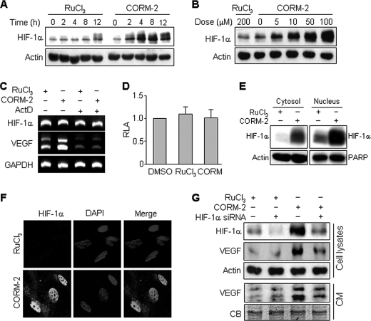
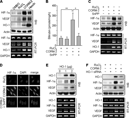
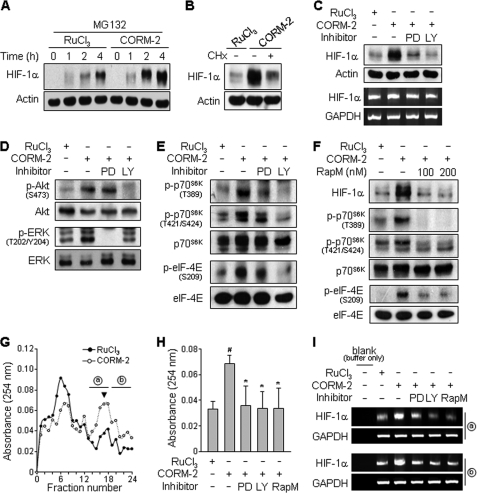
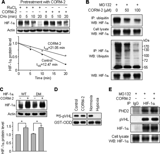
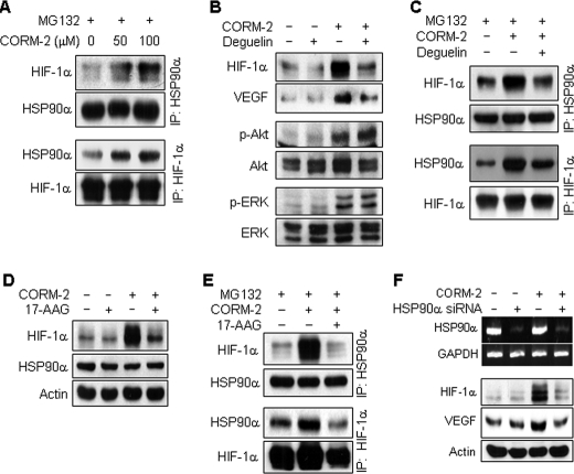
Carbon monoxide (CO) plays a significant role in vascular functions. We here examined the molecular mechanism by which CO regulates HIF-1 (hypoxia-inducible transcription factor-1)-dependent expression of vascular endothelial growth factor (VEGF), which is an important angiogenic factor. We found that astrocytes stimulated with CORM-2 (CO-releasing molecule) promoted angiogenesis by increasing VEGF expression and secretion. CORM-2 also induced HO-1 (hemeoxygenase-1) expression and increased nuclear HIF-1α protein level, without altering its promoter activity and mRNA level. VEGF expression was inhibited by treatment with HIF-1α siRNA and a hemeoxygenase inhibitor, indicating that CO stimulates VEGF expression via up-regulation of HIF-1α protein level, which is partially associated with HO-1 induction. CORM-2 activated the translational regulatory proteins p70(S6k) and eIF-4E as well as phosphorylating their upstream signal mediators Akt and ERK. These translational signal events and HIF-1α protein level were suppressed by inhibitors of phosphatidylinositol 3-kinase (PI3K), MEK, and mTOR, suggesting that the PI3K/Akt/mTOR and MEK/ERK pathways are involved in a translational increase in HIF-1α. In addition, CORM-2 also increased stability of the HIF-1α protein by suppressing its ubiquitination, without altering the proline hydroxylase-dependent HIF-1α degradation pathway. CORM-2 increased HIF-1α/HSP90α interaction, which is responsible for HIF-1α stabilization, and HSP90-specific inhibitors decreased this interaction, HIF-1α protein level, and VEGF expression. Furthermore, HSP90α knockdown suppressed CORM-2-induced increases in HIF-1α and VEGF protein levels. These results suggest that CO stimulates VEGF production by increasing HIF-1α protein level via two distinct mechanisms, translational stimulation and protein stabilization of HIF-1α.


Figures

FIGURE 1.
FIGURE 1.
CORM-2 up-regulates VEGF production via the augment of HIF-1α protein level. A and B, astrocytes were exposed to the indicated concentrations of RuCl3 and CORM-2 for various time periods (A) and for 8 h (B). HIF-1α and actin in cell lysates were analyzed by Western blotting. C, cells were pretreated with or without 1 μg/ml ActD for 30 min and exposed to RuCl3 (200 μm) or CORM-2 (100 μm) for 6 h. Cells were harvested, and total mRNAs were isolated. HIF-1α and VEGF mRNA levels were analyzed by RT-PCR. D, cells were transfected with pGL3-basic-pHIF-1α (0.4 μg) and pCMV-β-gal (0.4 μg) in 6-well plates. Transfected cells were incubated in fresh medium for 24 h and incubated with DMSO, RuCl3 (200 μm), and CORM-2 (100 μm) for 8 h. Cell lysates were obtained, and relative luciferase activity (RLA) was determined using a luminometer. E, cells were treated with RuCl3 (200 μm) or CORM-2 (100 μm) for 8 h. The cytosolic and nuclear fractions were isolated, and HIF-1α levels were detected by Western blotting. Actin and poly(ADP-ribose) polymerase (PARP) were used for internal controls. F, cells were treated with RuCl3 (200 μm) or CORM-2 (100 μm) for 8 h and fixed in 3.7% formaldehyde for 10 min. Cells were immunostained with an antibody against HIF-1α (green) and DAPI (blue). Fluorescent images were obtained using a confocal microscope. G, astrocytes were transfected with control or HIF-1α siRNA (100 nm), cultured in fresh medium for 40 h, and then treated with 200 μm RuCl3 or 100 μm CORM-2 for 8 h. Cells were harvested, and cell lysates were prepared. The cellular levels of HIF-1α and VEGF were analyzed by Western blotting. For CM preparation, cells were transfected with control or HIF-1α siRNA (100 nm) and cultured in fresh medium for 16 h, followed by treatment with 100 μm CORM-2 for 8 h. The media were replaced with FBS-free M199 and cultured for 24 h. CM were collected and concentrated through a centrifugal filter device. VEGF protein level in CM was determined by Western blotting. CB, a Coomassie Blue-stained protein band as an internal control. Error bars, S.D.
FIGURE 2.
FIGURE 2.
CORM-2-induced HIF-1α and VEGF expressions are regulated by HO-1 activity. A, astrocytes were treated with DMSO, RuCl3 (200 μm), CORM-2 (100 μm), and hemin (10 μm) for 8 h. B, astrocytes were transfected with mock or HO-1 expression vector and cultured in fresh medium for 48 h. HO activity was assayed as described under “Experimental Procedures.” Data shown are the mean ± S.D. (n = 3). *, p < 0.05; **, p < 0.01. C, astrocytes were treated with RuCl3 (200 μm) or CORM-2 (100 μm) for 8 h in the presence or absence of SnPP (50 μm). The protein and mRNA levels of HIF-1α, VEGF, and HO-1 were analyzed by Western blotting and RT-PCR. D, cells treated with RuCl3 (200 μm), CORM-2 (100 μm), or CORM-2 plus SnPP (50 μm) for 8 h were immunostained with an antibody against HIF-1α (green) and DAPI (blue). Fluorescent images were obtained by a confocal microscope. E, astrocytes were transfected with mock or HO-1 expression vector and cultured in fresh medium for 48 h. F, astrocytes were transfected with control or HO-1 siRNA (50 nm), cultured in fresh medium for 40 h, and then treated with 200 μm RuCl3 or 100 μm CORM-2 for 8 h. E and F, the protein and mRNA levels of HIF-1α, VEGF, and HO-1 were analyzed by Western blotting and RT-PCR. Error bars, S.D. WB, Western blot.
FIGURE 3.
FIGURE 3.
CORM-2 regulates HIF-1α translation via the PI3K- and MEK-dependent pathways. A, astrocytes were treated with RuCl3 (200 μm) or CORM-2 (100 μm) in the presence of MG132 (2.5 μm) for the indicated time periods, and cellular levels of HIF-1α and actin were determined by Western blotting. B, astrocytes were pretreated with or without 0.5 μg/ml CHx for 15 min and then incubated with RuCl3 (200 μm) or CORM-2 (100 μm) for 8 h. The cellular levels of HIF-1α were determined by Western blotting. C, cells were pretreated with 25 μm PD98059 (PD) or 10 μm LY294002 (LY) for 15 min and then treated with RuCl3 (200 μm) or CORM-2 (100 μm) for 8 h. HIF-1α protein and mRNA levels were analyzed by Western blotting and RT-PCR, respectively. D, cells were treated with RuCl3 (200 μm) or 100 μm CORM-2 in the presence or absence of 25 μm PD98059 or 10 μm LY294002 for 20 min. The cellular levels of phospho-Akt (p-Akt) and phospho-ERK (p-ERK) were determined by Western blotting. E and F, cells pretreated with 25 μm PD98059, 10 μm LY294002, or rapamycin (RapM; 100 and 200 nm) for 15 min and incubated with RuCl3 (200 μm) or 100 μm CORM-2 for 8 h. The levels of phospho-p70s6k (p-p70s6k), phospho-eIF-4B (p-eIF-4B), p70s6k, eIF-4B, and HIF-1α were determined by Western blot analyses. G–I, cells pretreated with 25 μm PD98059, 10 μm LY294002, or 100 nm rapamycin for 15 min and incubated with RuCl3 (200 μm) or 100 μm CORM-2 for 8 h. G, 20–50% sucrose gradients were separated in 24 fractions (0.2 ml), which were collected and monitored for absorbance at a 254-nm wavelength. Each absorbance was calculated by subtracting the absorbance from blank-containing lysis buffer without cell extract. The average absorbance from three independent experiments is shown. H, the average absorbance of fraction 17 (arrowhead in G) was monitored from three independent experiments. #, p < 0.005 versus untreated control; *, p < 0.05 versus CORM-2 alone. I, RNA was extracted from fractions (a, fractions 13–18; b, fractions 19–24) indicated in G, and HIF-1α and GAPDH mRNAs were detected by RT-PCR. Shown are representative data from three independent experiments.
FIGURE 4.
FIGURE 4.
CORM-2 increases HIF-1α protein stability. A, astrocytes were pretreated with CORM-2 (100 μm) for 8 h to increase HIF-1α protein level and further treated with RuCl3 (200 μm) or CORM-2 (100 μm) followed by CHx (0.5 μg/ml) treatment for the indicated time periods. The cellular levels of HIF-1α protein were determined by Western blot analysis. The relative protein levels of HIF-1α were calculated from the intensities of protein bands. B, astrocytes were treated with the indicated concentrations of CORM-2 combined with MG132 (10 μm) for 4 h, and cell extracts were subjected to immunoprecipitation (IP) using antibodies against ubiquitin and HIF-1α. Ubiquitinated HIF-1α was detected by immunoblotting (WB) these immunoprecipitates with antibodies against HIF-1α and ubiquitin. C, cells were transfected with wild-type (WT) HIF-1α or double-mutated (DM) (P402A/P564G) HIF-1α vector, cultured in fresh medium for 40 h, and then treated with or without CORM-2 (100 μm) for 8 h. HIF-1α protein level was assessed by Western blotting (n = 6). *, p < 0.05. D, cells were treated with or without CORM-2 (100 μm) for 4 h in the presence of MG132 (10 μm). Cell extracts were prepared and used for the PHD activity assay described under “Experimental Procedures.” Extracts prepared from cells cultured under normoxic and hypoxic conditions were used as controls. E, cells were treated with or without CORM-2 (100 μm) in the presence of MG132 (10 μm) for 4 h. Cell extracts were subjected to immunoprecipitation using an anti-HIF-1α antibody. Immunoprecipitates were separated by electrophoresis, and the protein levels of PHD2, pVHL, and HIF-1α were determined by Western blotting.
FIGURE 5.
FIGURE 5.
CORM-2 increases HIF-1α protein stability by promoting its interaction with HSP90α. A, astrocytes were treated with or without CORM-2 in the presence of MG132 (10 μm) for 4 h, and cell extracts were subjected to immunoprecipitation (IP) using antibodies against HSP90α and HIF-1α. Immunoprecipitates were separated by electrophoresis, and protein levels of HIF-1α and HSP90 were determined by Western blotting. B, astrocytes were pretreated with 100 nm deguelin for 15 min and incubated with or without CORM-2 (100 μm) for 8 h. The levels of HIF-1α, VEGF, phospho-Akt (p-Akt), and phospho-ERK (p-ERK) were determined by Western blot analyses. C, astrocytes pretreated with 100 nm deguelin for 15 min were incubated with or without CORM-2 (100 μm) in the presence of MG132 (10 μm) for 4 h. Cell extracts were subjected to immunoprecipitation using antibodies against HSP90α and HIF-1α. Immunoprecipitates were separated by electrophoresis, and the levels of HIF-1α or HSP90 were determined by Western blotting. D, cells pretreated with or without 10 μm 17-AAG for 15 min were incubated with or without CORM-2 (100 μm) for 8 h, and the levels of HIF-1α or HSP90 were determined by Western blotting. E, cells pretreated with 10 μm 17-AAG for 15 min and incubated with or without CORM-2 (100 μm) in the presence of MG132 (10 μm) for 4 h. Cell extracts were subjected to immunoprecipitation using an antibody against HSP90α or HIF-1α. Immunoprecipitates were analyzed by Western blotting with antibodies against HIF-1α and HSP90. F, cells were transfected with control or HSP90 siRNA (75 nm), cultured in fresh medium for 40 h, and then treated with or without CORM-2 (100 μm) for 8 h. HSP90α and GAPDH mRNA levels were determined by RT-PCR, and protein levels of HIF-1α and VEGF were detected by Western blot analyses.
FIGURE 6.
FIGURE 6.
CORM-2-treated astrocytes promote EC migration. A, astrocytes were pretreated with 25 μm PD98059 (PD), 10 μm LY294002 (LY), 50 μm SnPP (Sn), 10 μm 17-AAG (AA), or 100 nm deguelin (DG) in the lower chamber of Transwell plates for 15 min and treated with CORM-2 (COR; 100 μm) for 8 h. Cells were further cultured in fresh M199 containing 5% FBS for 20 h. HUVECs (1 × 10⁵ cells) were seeded on the upper chamber of Transwell plates. After a 4-h incubation, endothelial cell migration was detected by counting migrated endothelial cells after staining with hematoxylin and eosin using an optical microscope. Upper panel, microscopic image; lower panel, statistical data with the mean ± S.D. (n = 4). #, p < 0.001 versus untreated control; **, p < 0.01 versus CORM-2 alone. B, astrocytes pretreated with indicated inhibitors for 15 min and treated with CORM-2 for 8 h. Cells were further incubated with FBS-free DMEM for 24 h. Culture media were used for analyzing the levels of VEGF by Western blotting. CB, a Coomassie Blue-stained protein band as an internal control. Top, a representative immunoblot; bottom, statistical data with the mean ± S.D. (n = 3). #, p < 0.001 versus untreated control; *, p < 0.05; **, p < 0.01 versus CORM-2 alone.

Similar articles

Cited by

References

    1. Otterbein L. E., Zuckerbraun B. S., Haga M., Liu F., Song R., Usheva A., Stachulak C., Bodyak N., Smith R. N., Csizmadia E., Tyagi S., Akamatsu Y., Flavell R. J., Billiar T. R., Tzeng E., Bach F. H., Choi A. M., Soares M. P. (2003) Nat. Med. 9, 183–190 - PubMed
    1. Chauveau C., Bouchet D., Roussel J. C., Mathieu P., Braudeau C., Renaudin K., Tesson L., Soulillou J. P., Iyer S., Buelow R., Anegon I. (2002) Am. J. Transplant. 2, 581–592 - PubMed
    1. Foresti R., Bani-Hani M. G., Motterlini R. (2008) Intensive Care Med. 34, 649–658 - PubMed
    1. Brouard S., Otterbein L. E., Anrather J., Tobiasch E., Bach F. H., Choi A. M., Soares M. P. (2000) J. Exp. Med. 192, 1015–1026 - PMC - PubMed
    1. Tenhunen R., Marver H. S., Schmid R. (1968) Proc. Natl. Acad. Sci. U.S.A. 61, 748–755 - PMC - PubMed

Publication types

MeSH terms

Substances