. 2010 Oct 29;285(44):33923-9.
doi: 10.1074/jbc.M110.126896. Epub 2010 Aug 19.

ATP-dependent mechanism protects spectrin against glycation in human erythrocytes

Affiliations

ATP-dependent mechanism protects spectrin against glycation in human erythrocytes

Sumie Manno et al. J Biol Chem. .

Abstract

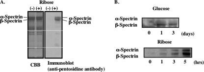
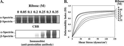
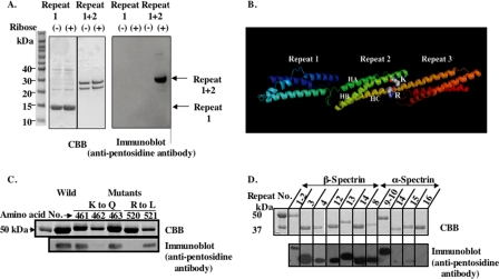
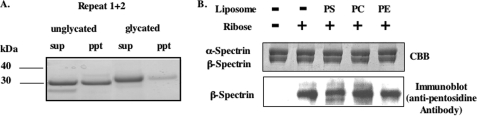
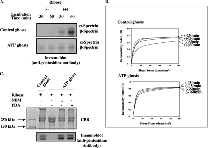
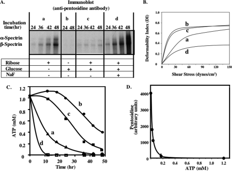
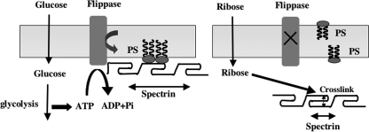
Human erythrocytes are continuously exposed to glucose, which reacts with the amino terminus of the β-chain of hemoglobin (Hb) to form glycated Hb, HbA1c, levels of which increase with the age of the circulating cell. In contrast to extensive insights into glycation of hemoglobin, little is known about glycation of erythrocyte membrane proteins. In the present study, we explored the conditions under which glucose and ribose can glycate spectrin, both on the intact membrane and in solution and the functional consequences of spectrin glycation. Although purified spectrin could be readily glycated, membrane-associated spectrin could be glycated only after ATP depletion and consequent translocation of phosphatidylserine (PS) from the inner to the outer lipid monolayer. Glycation of membrane-associated spectrin led to a marked decrease in membrane deformability. We further observed that only PS-binding spectrin repeats are glycated. We infer that the absence of glycation in situ is the consequence of the interaction of the target lysine and arginine residues with PS and thus is inaccessible for glycation. The reduced membrane deformability after glycation in the absence of ATP is likely the result of the inability of the glycated spectrin repeats to undergo the obligatory unfolding as a consequence of interhelix cross-links. We thus postulate that through the use of an ATP-driven phospholipid translocase (flippase), erythrocytes have evolved a protective mechanism against spectrin glycation and thus maintain their optimal membrane function during their long circulatory life span.


Figures

FIGURE 1.
FIGURE 1.
Glycation of erythrocyte ghosts membrane and purified spectrin. Erythrocyte ghosts membranes were prepared by hypotonic lysis and incubated with ribose at 37 °C for up to 6 h. A, glycated ghosts membrane proteins were separated on 9% SDS-polyacrylamide gel and stained with CBB (left panel); a Western blot of a replicate gel probed with the anti-pentosidine antibody is shown (right panel). B, purified spectrin dimers prepared from unglycated erythrocyte membranes were incubated with glucose (upper panel) or ribose (lower panel) for the indicated periods of days or hours, respectively.
FIGURE 2.
FIGURE 2.
Effects of ribose on spectrin glycation in erythrocytes and on membrane deformability. The membranes from erythrocytes incubated with the indicated concentrations of ribose at 37 °C for 48 h were prepared by hypotonic lysis and resealed in PBS. A, membrane proteins separated on 5% SDS-acrylamide gel and stained with CBB (upper panel) and blotted onto PVDF and probed with anti-pentosidine antibody (lower panel) are shown. B, membrane deformability of the ghosts was analyzed by ektacytometry.
FIGURE 3.
FIGURE 3.
Identification of pentosidine formation site of spectrin. Recombinant peptides of PS-binding repeats of α- and β-spectrin were prepared. A, repeat 1 and repeats 1 + 2 without GST were glycated and were separated on a one-dimensional 12% NuPAGE Novex BisTris Mini Gel in MES running buffer and blotted onto two PVDF membranes; one was stained with CBB (left panel) and the other probed with anti-pentosidine antibody (right panel). B, predicted ribbon model of repeats 1–3 of human erythroid β-spectrin based on the defined structure of brain βII-spectrin derived using software of Swiss Model are shown. HA, HB, and HC represent the helices of the triple-helical repeat 2. C, recombinant peptides of repeats 1 + 2 with indicated single-amino acid substitutions were glycated with ribose. They were separated by SDS-PAGE as indicated above. Gels were stained with CBB (upper panel), blotted onto PVDF, and probed with anti-pentosidine antibody (lower panel). D, various PS-binding and non-PS-binding repeats of α- and β-spectrin were glycated with ribose. They were separated by SDS-PAGE as above. Gels were stained with CBB (upper panel), blotted onto PVDF, and probed with anti-pentosidine antibody (lower panel).
FIGURE 4.
FIGURE 4.
Effect of glycation on binding of spectrin to PS-liposomes and extent of glycation of spectrin in the presence of PS-liposomes. A, repeats 1 + 2 were glycated with ribose. Glycated or unglycated repeat 1 + 2 polypeptides were incubated with PS-liposomes at room temperature for 60 min. The liposomes were then collected by centrifugation. The supernatants (sup) and liposome pellets (ppt) were analyzed by SDS-PAGE and stained with CBB. B, purified spectrin was incubated with ribose in the presence of PS-, PC-, and PE-liposomes (molar ratio of 7 to spectrin dimer) at 37 °C for 8 h. Pentosidine formation of spectrin molecules was analyzed by SDS-PAGE. Gels were stained with CBB (upper panel), blotted onto PVDF, and probed with anti-pentosidine antibody (lower panel).
FIGURE 5.
FIGURE 5.
Glycation of ATP ghosts. The ghosts were prepared by lysis of cells in the presence or absence of ATP and resealed as described under “Methods.” These ghosts were glycated with ribose at 37 °C for up to 60 min. A, membrane proteins were separated on 5% SDS-PAGE followed by immunoblotting with anti-pentosidine antibody. B, membrane deformability of these ghosts was measured by the ektacytometry. C, flippase inhibitor, PDA or N-ethylmaleimide (NEM), was incorporated into ATP ghosts. Pentosidine formation of spectrin molecules was analyzed by SDS-PAGE. Gels were stained with CBB, blotted onto PVDF, and probed with anti-pentosidine antibody.
FIGURE 6.
FIGURE 6.
Effects of ATP on pentosidine formation and membrane deformability. Erythrocytes were incubated for up to 48 h with ribose alone (a), glucose alone (b), a mixture of ribose and glucose (c), or the same mixture with NaF (d). Ghosts were prepared from these erythrocytes by hypotonic lysis and resealed in isotonic buffer. A, ghost proteins were separated on 5% SDS-PAGE and blotted onto PVDF membranes and probed with anti-pentosidine antibody. B, deformability of these ghosts was measured in the ektacytometer. C, measured ATP concentrations were plotted against incubation time. D, pentosidine formation was plotted against intracellular ATP concentrations.
FIGURE 7.
FIGURE 7.
Working model for glycation of spectrin induced by either glucose or ribose in human erythrocytes. In normal erythrocytes, glycolysis-derived ATP driving the ATP-dependent flippase maintains PS in the inner monolayer, enabling the PS-binding spectrin repeats to be anchored in the bilayer (left panel). In the absence of ATP, PS is translocated to the outer monolayer, enabling the dissociation of PS-binding repeats from the membrane and thereby making the lysine and arginine residues accessible for glycation.

References

    1. Bierhaus A., Hofmann M. A., Ziegler R., Nawroth P. P. (1998) Cardiovasc. Res. 37, 586–600 - PubMed
    1. Sell D. R., Monnier V. M. (1989) J. Biol. Chem. 264, 21597–21602 - PubMed
    1. Grandhee S. K., Monnier V. M. (1991) J. Biol. Chem. 266, 11649–11653 - PubMed
    1. Bunn H. F., Gabbay K. H., Gallop P. M. (1978) Science 200, 21–27 - PubMed
    1. Mohandas N., Gallagher P. G. (2008) Blood 112, 3939–3948 - PMC - PubMed

Publication types

MeSH terms

LinkOut - more resources