. 2010 Oct 29;285(44):33602-13.
doi: 10.1074/jbc.M110.135640. Epub 2010 Aug 19.

The obligatory intestinal folate transporter PCFT (SLC46A1) is regulated by nuclear respiratory factor 1

Affiliations

The obligatory intestinal folate transporter PCFT (SLC46A1) is regulated by nuclear respiratory factor 1

Nitzan Gonen et al. J Biol Chem. .

Abstract

Folates are essential vitamins that play a key role as one-carbon donors in a spectrum of biosynthetic pathways including RNA and DNA synthesis. The proton-coupled folate transporter (PCFT/SLC46A1) mediates obligatory intestinal folate absorption. Loss-of-function mutations in PCFT result in hereditary folate malabsorption, an autosomal recessive disorder characterized by very low folate levels in the blood and cerebrospinal fluid. Hereditary folate malabsorption manifests within the first months after birth with anemia, immune deficiency, and neurological deficits. Here we studied the role of inducible trans-activators of PCFT gene expression. Bioinformatics identified three putative nuclear respiratory factor 1 (NRF-1) binding sites in the minimal promoter. The following evidence establish that PCFT is an NRF-1-responsive gene; electrophoretic mobility shift assay showed NRF-1 binding to native but not mutant NRF-1 sites, whereas antibody-mediated supershift analysis and chromatin immunoprecipitation revealed NRF-1 binding to its consensus sites within the PCFT promoter. Moreover, mutational inactivation of individual or all NRF-1 binding sites resulted in 40-60% decrease in luciferase reporter activity. Consistently, overexpression of NRF-1 or a constitutively active NRF-1 VP-16 construct resulted in increased reporter activity and PCFT mRNA levels. Conversely, introduction of a dominant-negative NRF-1 construct markedly repressed reporter activity and PCFT mRNA levels; likewise, introduction of NRF-1 siRNA duplexes to cells resulted in decreased PCFT transcript levels. Moreover, NRF-1 silencing down-regulated genes encoding for key folate transporters and enzymes in folate metabolism. These novel findings identify NRF-1 as a major inducible transcriptional regulator of PCFT gene expression. The implications of this linkage between folate transport and metabolism with mitochondria biogenesis and respiration are discussed.


Figures

FIGURE 1.
FIGURE 1.
Identification of three putative NRF-1 binding sites in the human PCFT promoter. Alignment of the 5′ region of the human PCFT promoter containing 296 bp upstream to the ATG (23, 24) with that of the following mammals: monkey, cow, mouse, and rat. Putative NRF-1 binding sites termed NRF-1 A, B, and C are denoted by open squares (the dark box in NRF-1A represents the highest bioinformatic score). The beginning of the 5′-UTR is marked by the arrow. Nucleotide conservation is indicated by the asterisks in all mammalian organisms studied.
FIGURE 2.
FIGURE 2.
NRF-1 binds and activates the human PCFT promoter. A, EMSA was performed using nuclear extract from HeLa cells and oligonucleotides containing the authentic sequence of NRF-1 sites from the minimal PCFT promoter region. DNA-protein complex a and the high molecular weight complex formed using antibody-dependent supershift analysis (complex b) were resolved by electrophoresis on non-denaturing polyacrylamide gels and examined by a phosphorimager as detailed under “Experimental Procedures.” Molar excess competitions (250-fold) were performed with nonradioactive oligonucleotides. The sequence of the consensus and mutant NRF-1 oligonucleotides is indicated in the lower left side of the panel. The core NRF-1 binding site is underlined, and inactivating mutations in the core consensus are depicted in bold. B, chromatin immunoprecipitation was performed using mouse IgG, rabbit IgG as well as polymerase II (PolII) and NRF-1-specific antibodies. Immunoprecipitation (Ip) in the upper panel corresponds to PCR targeting the PCFT promoter region −130 to +90 (220 bp). Ip in the lower panel corresponds to PCR targeting a 174-bp region of an intergenic genomic DNA sequence from chromosome 12 (negative control). C, luciferase reporter gene assay in HeLa cells following transient co-transfection with pRL-O Renilla plasmid and the various pGL3-271 bp PCFT promoter constructs harboring point mutations (indicated by X) of the designated NRF-1 sites (black rectangles). Results are presented as a luciferase/Renilla ratio (%), normalized to the pGL3-271-bp vector assigned 100% activity. Results are the mean ± S.D of three independent experiments performed in duplicates.
FIGURE 3.
FIGURE 3.
Luciferase reporter assay reveals that PCFT promoter activity is modulated by NRF-1 levels. A–C, HeLa cells were transiently co-transfected with plasmids: 1) pRL-O Renilla; 2) pGL3-271 bp PCFT promoter construct; and 3) various amounts (ng) of NRF-1 WT (A), NRF-1 VP16 (B), or NRF-1 DN (C). Luciferase activity was then determined as detailed under “Experimental Procedures.” Results are presented as a luciferase/Renilla ratio (%), normalized to the pGL3-271-bp construct along with the corresponding amount of pCDNA empty vector assigned 100% activity. D, luciferase reporter assay in HeLa cells following co-transfection with plasmids: 1) pRL-O Renilla plasmid; 2) 2.5 ng of NRF-1 WT or NRF-1 DN; 3) PCFT promoter constructs pGL3-3.1 kb, -271 bp, or -157 bp. Results are presented as a luciferase/Renilla ratio (%), normalized to the corresponding PCFT promoter construct along with the 2.5 ng of pCDNA empty vector assigned 100% activity. Results shown are the mean ± S.D of three independent experiments performed in duplicates.
FIGURE 4.
FIGURE 4.
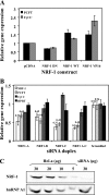
NRF-1 regulates PCFT mRNA levels. A, HeLa cells were stably transfected with one of the following expression vectors: pCDNA3, NRF-1 DN, NRF-1 WT, or NRF-1 VP16 and a stable population was established by drug selection using G418 or hygromycin. PCFT and cytochrome c (CytC) mRNA levels were determined using real time PCR. Gene expression was normalized to β2-microglobulin mRNA levels. Results are presented as relative expression levels of PCFT or cytochrome c mRNA when compared with cells transfected with the empty pCDNA3 vector, which was assigned a value of 1. B, NRF-1 RNA interference leads to down-regulation of PCFT mRNA; HeLa cells were transfected with each of three different siRNA duplexes that targeted NRF-1 or a combination of all three. For a negative control, HeLa cells were transfected with scrambled siRNA that was not directed to any gene. The mRNA expression level of PCFT, cytochrome c, NRF-1, and hypoxanthine-guanine phosphoribosyltransferase (HPRT) were determined using real time PCR. Gene expression was normalized to β2-microglobulin mRNA. Results are presented relative to the expression of the above genes in a mock transfection, assigned a value of 1. Results are the means of three independent experiments ± S.D. C, Western blot analysis of NRF-1 protein levels following NRF-1 silencing via siRNA. HeLa cells were transfected with NRF-1 C siRNA duplex and nuclear proteins were isolated. Different amounts of nuclear proteins from HeLa cells (5, 10, 20, and 30 μg) as well as 30 μg of nuclear proteins from NRF-1-silenced cells were resolved by electrophoresis on 10% polyacrylamide gels under denaturing conditions. NRF-1 protein levels were determined using an anti-NRF-1 antibody. The protein level of the nuclear splicing factor heterogeneous nuclear ribonucleoprotein (hnRNP) A1 was used for estimation of equal loading.
FIGURE 5.
FIGURE 5.
Bioinformatic analysis of consensus NRF-1 binding sites in the promoters of genes encoding for folate transporters and enzymes in the folate metabolic pathway. The bioinformatic program Genomatics was used to predict the presence of potential NRF-1 binding sites in the 2-kb promoter region as well as the 5′ UTRs of the above genes, the expression of which was decreased following NRF-1 knockdown. The putative NRF-1 binding sites are denoted by solid rectangles. The position of the 5′-most nucleotide of each putative NRF-1 binding site is shown above the solid rectangle.
FIGURE 6.
FIGURE 6.
PGC-1α does not collaborate with NRF-1 in the regulation of PCFT gene expression. HeLa cells were co-transfected with plasmids: 1) pRL-O Renilla; 2) pGL3-271 bp PCFT promoter construct; 3) 2.5 or 25 ng of one of the following plasmids: NRF-1 WT, NRF-1 VP16, PGC-1α, or a combination of them, as indicated in the figure. Luciferase reporter activity was then determined. Results are presented as a luciferase/Renilla ratio (%), normalized to the pGL3-271 bp construct along with the appropriate amount of pCDNA empty vector assigned 100% activity. Results are the mean ± S.D of three independent experiments performed in duplicates.

Similar articles

Cited by

References

    1. Appling D. R. (1991) FASEB J. 5, 2645–2651 - PubMed
    1. Stockstad E. L. R. (1990) Folic Acid Metabolism in Health and Disease, pp. 1–21, Wiley-Liss, New York
    1. Assaraf Y. G. (2007) Cancer Metastasis Rev. 26, 153–181 - PubMed
    1. Dixon K. H., Lanpher B. C., Chiu J., Kelley K., Cowan K. H. (1994) J. Biol. Chem. 269, 17–20 - PubMed
    1. Matherly L. H., Hou Z., Deng Y. (2007) Cancer Metastasis Rev. 26, 111–128 - PubMed

LinkOut - more resources