. 2010 Dec 2;116(23):5021-31.
doi: 10.1182/blood-2010-03-276964. Epub 2010 Aug 19.

Monocyte-bound PF4 in the pathogenesis of heparin-induced thrombocytopenia

Affiliations

Monocyte-bound PF4 in the pathogenesis of heparin-induced thrombocytopenia

Lubica Rauova et al. Blood. .

Abstract

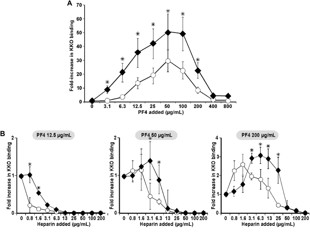
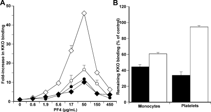
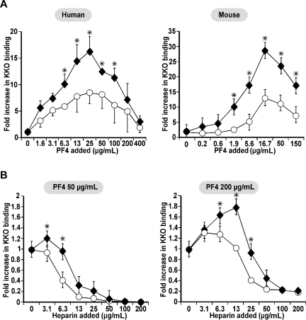
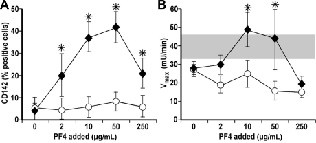
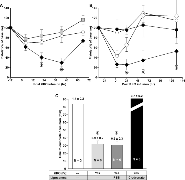
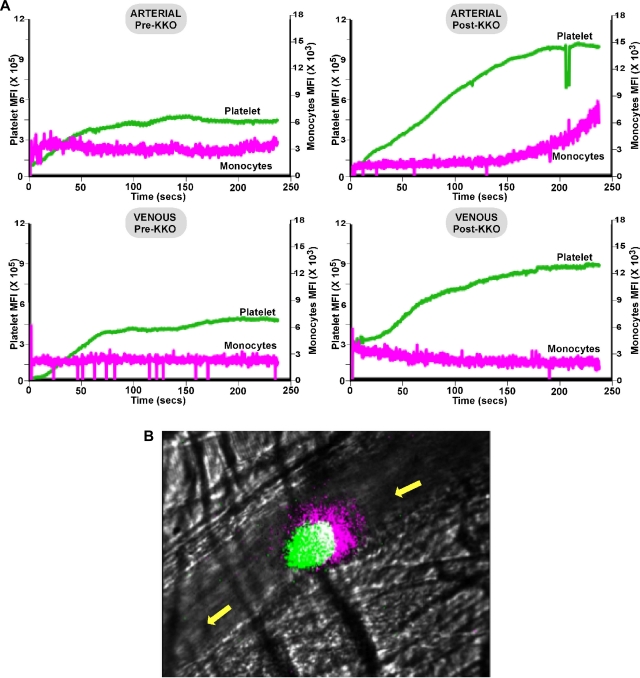
Heparin-induced thrombocytopenia (HIT) is a life- and limb-threatening thrombotic disorder that develops after exposure to heparin, often in the setting of inflammation. We have shown previously that HIT is associated with antibodies to complexes that form between platelet factor 4 and glycosaminoglycan (GAG) side chains on the surface of platelets. However, thrombosis can occur in the absence of thrombocytopenia. We now show that platelet factor 4 binds to monocytes and forms antigenic complexes with their surface GAG side chains more efficiently than on platelets likely due to differences in GAG composition. Binding to monocytes is enhanced when the cells are activated by endotoxin. Monocyte accumulation within developing arteriolar thrombi was visualized by situ microscopy. Monocyte depletion or inactivation in vivo attenuates thrombus formation induced by photochemical injury of the carotid artery in a modified murine model of HIT while paradoxically exacerbating thrombocytopenia. These studies demonstrate a previously unappreciated role for monocytes in the pathogenesis of arterial thrombosis in HIT and suggest that therapies targeting these cells might provide an alternative approach to help limit thrombosis in this and possibly other thrombotic disorders that occur in the setting of inflammation.


Figures

Figure 1
Figure 1
Surface PF4/GAG antigenicity on monocytes compared with platelets. (A) PF4/GAG complexes detected using the HIT-like monoclonal antibody KKO on human monocytes (♦) compared with simultaneously studied human platelets from the same blood sample (○) and (B) in the presence of increasing concentration of heparin (0.8-200 μg/mL ≈ 0.17-42.4 USP/mL) at 3 fixed PF4 concentrations: 12.5 μg/mL (left), 50 μg/mL (center), and 200 μg/mL (right). The graphs show the fold-increase in mean fluorescence intensity (MFI) of antibody binding in the presence of the concentrations of PF4 and heparin noted compared with MFI in its absence after the results were normalized for size based on equalizing the background MFI of monocytes and platelets. N ≥ 3 experiments, each done in duplicate. The mean ± 1 SE is shown. *P < .05, comparing the fold increase in MFI for monocytes to platelets at a specific PF4 concentration.
Figure 2
Figure 2
Studies of surface GAGs in KKO binding to cell surface. (A) KKO binding to monocytes (diamonds) and to platelets (circles) at increasing concentrations of PF4 as in Figure 1A but with either the vehicle DMSO (open symbols) or in the presence of 10 μM surfen (closed symbols). N = 5 experiments, each done in duplicate. The mean ± 1 SE is shown. (B) Monocytes and platelets pretreated with either chondroitinase ABC alone (■) or heparinase alone (□). Residual KKO binding compared with control (untreated) cells at the optimal PF4 concentration (50 μg/mL) is shown. N = 3 experiments, each done in duplicate. The mean ± 1 SE is shown.
Figure 3
Figure 3
Surface KKO binding on monocytes compared with macrophages. (A) Human (left) and mouse (right) studies of relative KKO binding as in Figure 1A to cultured unstimulated monocytes (□) and concurrently studied LPS-stimulated monocytes (♦). The murine cells were isolated from a mPF4KO mouse. N ≥ 3, each in duplicate. Mean ± 1 SE are shown. *P < .05 for relative binding to stimulated cells versus resting cells. (B) mPF4KO mice cultured, unstimulated monocyte (○), and concurrently studied LPS-stimulated monocytes (♦) at 2 different PF4 concentrations and different heparin concentrations as in Figure 1B. N ≥ 3, each in duplicate. Mean ± 1 SE are shown. *P < .05 for relative binding to stimulated cells versus unstimulated cells.
Figure 4
Figure 4
Expression and activity of TF on monocytes in the presence of PF4 and KKO. (A) Expression of TF on monocytes at different PF4 concentrations in the absence (○) or presence of KKO (♦). N ≥ 3, each in duplicate. Mean ± 1 SE are shown. *P < .05 for relative binding in the absence versus presence of KKO. (B) Same as in panel A but for TF activity. Gray bar represents the range of TF activity after LPS stimulation. N ≥ 3, each in duplicate. Mean ± 1 SE are shown. *P < .05 for relative binding in the absence versus presence of KKO.
Figure 5
Figure 5
Effect of monocyte depletion on platelet count and thrombosis in the HIT model. (A) Platelet counts after induction of HIT in the FcγRIIA+/hPF4+ mice model. Clodronate- or PBS-laden liposomes were injected intravenously. Twelve hours later (Time 0), KKO 2.5 mg/kg was administered intraperitoneally. The first platelet count was measured 4 hours later. Animals receiving no liposomes (○) or PBS-laden liposomes (▩) had modest thrombocytopenia compared with the same mice receiving clodronate-laden liposomes (♦). N ≥ 3, each in duplicate. Mean ± 1 SE are shown. *P < .05 for platelet drop after KKO in mice with depleted monocytes versus control mice. (B) Same as panel A but animals received NaCl (open symbols) or GdCl3 (black symbols) 32 hours prior to the KKO 10 mg/kg intraperitoneal injection (diamonds) or controls without KKO (circles). N ≥ 3, each in duplicate. Mean ± 1 SE are shown. *P < .05 for platelet drop in mice treated with GdCl3 followed by KKO versus mice treated with NaCl followed by KKO. (C) Times to complete occlusion in a photochemical carotid artery model in FcγRIIA+/hPF4+ mice under conditions noted in the figure are shown as mean ± 1 SE. The platelet counts at the time of study are indicated above each bar and is per 109/mL. N values are noted in the figures. *P < .001 for time to occlusion relative to mice not receiving either KKO or liposomes.
Figure 6
Figure 6
In situ laser injury studies in the murine HIT model. (A) Shown are cumulative information for platelets (green) and monocytes (pink) at a site of induced injury for ≥ 10 injuries per condition within the arterioles and venules and pre- and post-KKO as indicated. (B) Same as in panel A but still from video at 4 minutes from an arteriole injury showing the accumulation of CD115+-monocytes and/or microparticles at the upstream end of a growing thrombus. Platelets appear green, monocytes pink, and the overlap white. The yellow arrow denotes the direction of blood flow.

Similar articles

Cited by

References

    1. Loscalzo J, Melnick B, Handin RI. The interaction of platelet factor four and glycosaminoglycans. Arch Biochem Biophys. 1985;240(1):446–455. - PubMed
    1. George JN, Onofre AR. Human platelet surface binding of endogenous secreted factor VIII-von Willebrand factor and platelet factor 4. Blood. 1982;59(1):194–197. - PubMed
    1. Rao AK, Niewiarowski S, James P, et al. Effect of heparin on the in vivo release and clearance of human platelet factor 4. Blood. 1983;61(6):1208–1214. - PubMed
    1. Amiral J, Bridey F, Dreyfus M, et al. Platelet factor 4 complexed to heparin is the target for antibodies generated in heparin-induced thrombocytopenia. Thromb Haemost. 1992;68(1):95–96. - PubMed
    1. Greinacher A, Potzsch B, Amiral J, Dummel V, Eichner A, Mueller-Eckhardt C. Heparin-associated thrombocytopenia: isolation of the antibody and characterization of a multimolecular PF4-heparin complex as the major antigen. Thromb Haemost. 1994;71(2):247–251. - PubMed

Publication types