

Alveolar macrophages stimulate enhanced cytokine production by pulmonary CD4+ T-lymphocytes in an exacerbation of murine chronic asthma
- PMID: 20724599
- PMCID: PMC2947263
- DOI: 10.2353/ajpath.2010.100019
Alveolar macrophages stimulate enhanced cytokine production by pulmonary CD4+ T-lymphocytes in an exacerbation of murine chronic asthma
Abstract
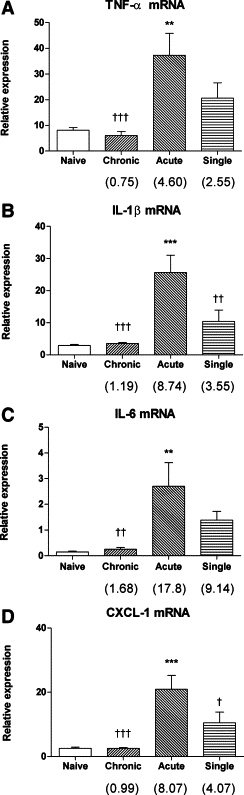
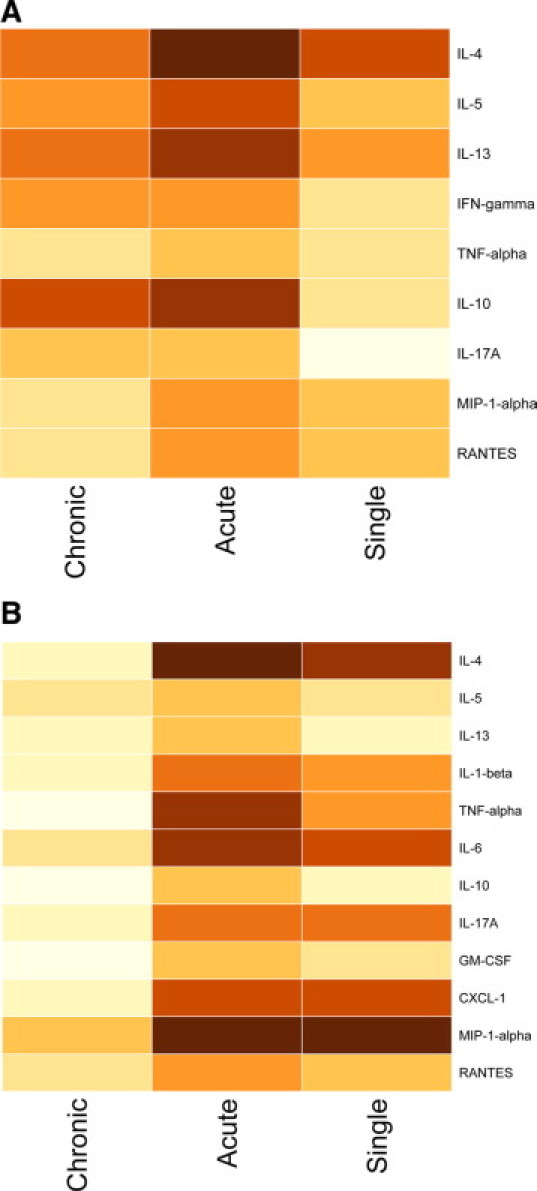
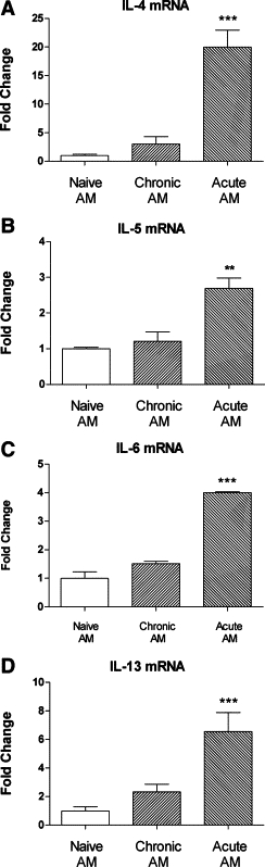
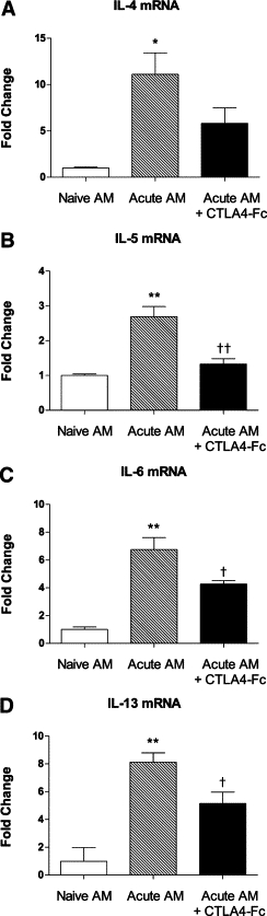
The mechanisms underlying the exaggerated distal airway inflammation and hyperresponsiveness that characterize acute exacerbations of asthma are largely unknown. Using BALB/c mouse experimental models, we demonstrated a potentially important role for alveolar macrophages (AM) in the development of an allergen-induced exacerbation of asthma. To induce features of airway inflammation and remodeling characteristic of mild chronic asthma, animals were systemically sensitized and exposed to low mass concentrations (≈3 mg/m(3)) of aerosolized ovalbumin for 30 minutes per day, 3 days per week, for 4 weeks. A subsequent single moderate-level challenge (≈30 mg/m(3)) was used to trigger an acute exacerbation. In chronically challenged animals, cytokine expression by AM was not increased, whereas after an acute exacerbation, AM exhibited significantly enhanced expression of proinflammatory cytokines, including interleukin (IL) 1β, IL-6, CXCL-1, and tumor necrosis factor α. In parallel, there was a marked increase in the expression of several cytokines by CD4(+) T-lymphocytes, notably the Th2 cytokines IL-4 and IL-13. Importantly, AM from an acute exacerbation stimulated the expression of Th2 cytokines when cocultured with CD4(+) cells from chronically challenged animals, and their ability to do so was significantly greater than AM from either chronically challenged or naïve controls. Stimulation was partly dependent on interactions involving CD80/86. We conclude that in an acute exacerbation of asthma, enhanced cytokine expression by AM may play a critical role in triggering increased expression of cytokines by pulmonary CD4(+) T-lymphocytes.
Figures
References
-
- Holgate ST. Exacerbations: the asthma paradox. Am J Respir Crit Care Med. 2005;172:941–943. - PubMed
-
- Martin RJ. Therapeutic significance of distal airway inflammation in asthma. J Allergy Clin Immunol. 2002;109:S447–S460. - PubMed
-
- Robinson DS, Hamid Q, Ying S, Tsicopoulos A, Barkans J, Bentley AM, Corrigan C, Durham SR, Kay AB. Predominant TH2-like bronchoalveolar T-lymphocyte population in atopic asthma. N Engl J Med. 1992;326:298–304. - PubMed
Publication types
MeSH terms
Substances
LinkOut - more resources
Full Text Sources
Medical
Molecular Biology Databases
Research Materials
