. 2010 Oct;177(4):1848-60.
doi: 10.2353/ajpath.2010.091006. Epub 2010 Aug 19.

PrPC, the cellular isoform of the human prion protein, is a novel biomarker of HIV-associated neurocognitive impairment and mediates neuroinflammation

Affiliations

PrPC, the cellular isoform of the human prion protein, is a novel biomarker of HIV-associated neurocognitive impairment and mediates neuroinflammation

Toni K Roberts et al. Am J Pathol. 2010 Oct.

Abstract

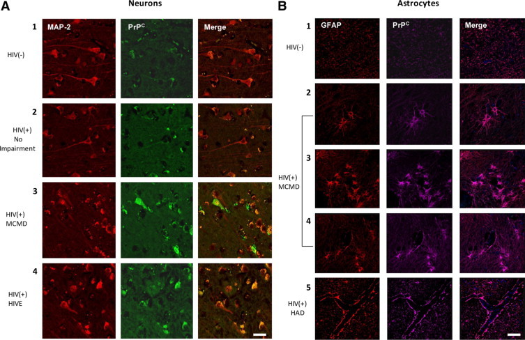
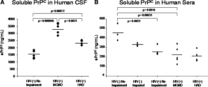
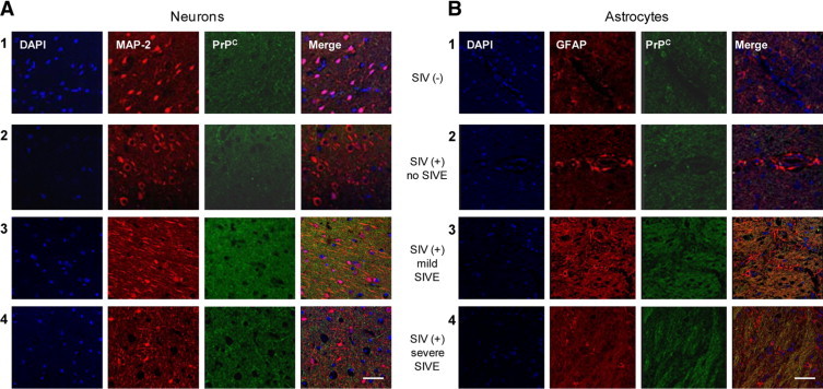
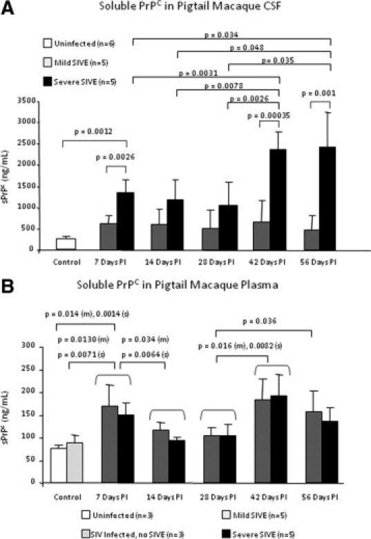
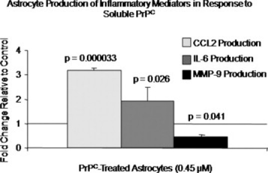
Of the 33 million people infected with the human immunodeficiency virus (HIV) worldwide, 40-60% of individuals will eventually develop neurocognitive sequelae that can be attributed to the presence of HIV-1 in the central nervous system (CNS) and its associated neuroinflammation despite antiretroviral therapy. PrP(C) (protease resistant protein, cellular isoform) is the nonpathological cellular isoform of the human prion protein that participates in many physiological processes that are disrupted during HIV-1 infection. However, its role in HIV-1 CNS disease is unknown. We demonstrate that PrP(C) is significantly increased in both the CNS of HIV-1-infected individuals with neurocognitive impairment and in SIV-infected macaques with encephalitis. PrP(C) is released into the cerebrospinal fluid, and its levels correlate with CNS compromise, suggesting it is a biomarker of HIV-associated neurocognitive impairment. We show that the chemokine (c-c Motif) Ligand-2 (CCL2) increases PrP(C) release from CNS cells, while HIV-1 infection alters PrP(C) release from peripheral blood mononuclear cells. Soluble PrP(C) mediates neuroinflammation by inducing astrocyte production of both CCL2 and interleukin 6. This report presents the first evidence that PrP(C) dysregulation occurs in cognitively impaired HIV-1-infected individuals and that PrP(C) participates in the pathogenesis of HIV-1-associated CNS disease.


Figures

Figure 1
Figure 1
PrPC is increased significantly in the brain of individuals with HIV-1-associated neurocognitive impairment. Frontal cerebral cortex was obtained at time of autopsy from individuals who were HIV negative, unimpaired (HIV−), n = 4; HIV-infected, unimpaired (HIV+), n = 2; HIV-infected with minor motor cognitive disorder (MCMD), n = 4; HIV-infected with HIV-associated dementia (HAD), n = 2; and HIV-infected with HIV encephalitis (HIVE), n = 2. Immunohistochemistry was performed on paraffin-embedded tissue sections and was imaged using confocal microscopy. A: Neuronal expression of PrPC (FITC-green) was evaluated using MAP-2 (Cy3-red; neuronal marker). PrPC was increased in individuals with MCMD and HIVE relative to uninfected (panel 1) and infected individuals (panel 2) who were unimpaired. B: Astrocyte expression of PrPC was indicated by GFAP (Cy3-red; astrocytic marker) and PrPC (Cy5-magenta) colocalization. PrPC expression was elevated in astrocytes showing HIV-mediated pathology including hypertrophy (panel 2), proliferation (panel 3), and extensive process formation (panel 4) in individuals with HAD as compared to uninfected individuals (panel 1). Astrocyte PrPC was also increased at the blood–brain barrier where astrocyte foot processes contact the endothelium in individuals with HAD (panel 5), relative to levels in uninfected individuals (panel 1). PrPC was consistently expressed in all tissues.
Figure 2
Figure 2
CSF soluble PrPC levels predict neurocognitive impairment in HIV-1–infected individuals. CSF was obtained from individuals diagnosed as HIV-1–infected, neurocognitively normal; HIV-1–infected with minor motor cognitive disorder (MCMD); HIV-1–infected with HIV-associated dementia (HAD); uninfected, neurocognitively normal; and uninfected with neuropsychiatric impairment. Soluble PrPC (sPrPC) was evaluated by ELISA. A: CSF levels of sPrPC were elevated in individuals with HIV-associated neurocognitive impairment (MCMD and HAD) relative to infected individuals without neurocognitive impairment. Individuals with MCMD had higher CSF sPrPC levels than those with HAD, likely due to loss of neuropil. Soluble PrPC mean values: 1500.98 ng/ml (HIV-1–infected, unimpaired); 3348.07 ng/ml (HIV-1–infected, MCMD); 2372.81 ng/ml (HIV-1–infected, HAD). B: Sera was obtained from uninfected, unimpaired individuals; uninfected, neurocognitively impaired individuals; HIV-1–infected, unimpaired individuals; HIV-1–infected individuals with MCMD; and HIV-1–infected individuals with HAD, and was evaluated for sPrPC by ELISA. Neurocognitively unimpaired and impaired individuals who were infected with HIV-1 had lower levels of sPrPC in their sera than uninfected individuals. However, there was no difference in sera sPrPC among HIV-1–infected individuals who were cognitively normal or cognitively impaired. Thus CSF but not sera sPrPC may be a specific biomarker of neurocognitive impairment and CNS dysfunction in individuals with HIV-associated neurocognitive impairment. Soluble PrPC mean values: 455.33 ng/ml (uninfected, unimpaired); 329.08 ng/ml (uninfected, neurocognitively impaired); 254.81 ng/ml (HIV-1–infected, unimpaired); 220.92 ng/ml (HIV-1–infected, MCMD); and 202.70 ng/ml (HIV-1–infected, HAD).
Figure 3
Figure 3
CNS PrPC is increased in pigtail macaques with SIV encephalitis. Brain sections from two uninfected pigtail macaques, one SIV-infected macaque without encephalitis, and three SIV-infected macaques with mild and three with severe SIV encephalitis (SIVE) were evaluated by immunohistochemistry using confocal microscopy. A: Neuronal expression of PrPC (FITC-green) was evaluated using MAP2 (Cy3-red; neuronal marker). B: Astrocyte expression of PrPC was indicated by GFAP (Cy3-red; astrocytic marker) and PrPC (FITC-green) colocalization. PrPC expression was elevated in neurons and astrocytes in animals with encephalitis (panels 3 and 4) relative to uninfected animals (panel 1) and animals infected with SIV without encephalitis (panel 2). There was no detectable difference in PrPC expression between mildly and severely encephalitis animals. PrPC was consistently elevated in all encephalitis tissue examined.
Figure 4
Figure 4
CSF soluble PrPC levels correlate with severity of encephalitis in the pigtail macaque model of neuroAIDS. CSF and plasma was obtained once per week after infection with SIV until the time of sacrifice at 56 days postinoculation (PI). Soluble PrPC was evaluated by ELISA. A: The CSF from animals that developed severe SIVE contained higher levels of sPrPC than the CSF of those developing mild SIVE with a pronounced difference seen in late stages of disease. The increased levels of CSF sPrPC in severely encephalitic animals at days 42 and 56 PI correlated with increased CSF viral RNA and CCL2 levels. B: The plasma from SIV-infected encephalitic pigtail macaques (m = mild, s = severe) contained higher levels of sPrPC than the plasma from uninfected and infected animals without encephalitis at time points associated with high CNS viral load and inflammation. There was no difference in plasma sPrPC levels in animals that developed mild and severe encephalitis. Increased sPrPC at 7 days PI corresponded with high CNS viral activity and CSF CCL2 levels, followed by an antiviral and immunosuppressive response between 14–28 days PI and a concomitant drop in sPrPC levels. At 42 days PI, concordant with recrudescence of neuroinflammation and rebound of CNS viral load, CSF and plasma levels of sPrPC rose. CSF but not plasma sPrPC predicts severity of encephalitis in SIV-infected pigtail macaques.
Figure 5
Figure 5
Soluble PrPC alters the production of inflammatory mediators by astrocytes. Astrocytes were treated with 0.45 μmol/L recombinant human full-length PrPC (recPrPC) for 6 hours, followed by collection of media and evaluation by ELISA in three independent experiments. Astrocyte production of CCL2, IL-6, and MMP-9 in response to recPrPC was evaluated relative to vehicle-treated cells and is reported as fold change. Astrocytes secreted CCL2 and IL-6 in response to recPrPC, whereas their production of MMP-9 was reduced below baseline. These findings indicate that sPrPC modulates the inflammatory response of astrocytes and contributes to HIV-mediated neuroinflammation.

Similar articles

Cited by

References

    1. UNAIDS . Report on the global AIDS epidemic 2008. World Health Organization; Geneva: 2008.
    1. Tozzi V, Balestra P, Lorenzini P, Bellagamba R, Galgani S, Corpolongo A, Vlassi C, Larussa D, Zaccarelli M, Noto P, Visco-Comandini U, Giulianelli M, Ippolito G, Antinori A, Narciso P. Prevalence and risk factors for human immunodeficiency virus-associated neurocognitive impairment, 1996 to 2002: results from an urban observational cohort. J Neurovirol. 2005;11:265–273. - PubMed
    1. Antinori A, Arendt G, Becker JT, Brew BJ, Byrd DA, Cherner M, Clifford DB, Cinque P, Epstein LG, Goodkin K, Gisslen M, Grant I, Heaton RK, Joseph J, Marder K, Marra CM, McArthur JC, Nunn M, Price RW, Pulliam L, Robertson KR, Sacktor N, Valcour V, Wojna VE. Updated research nosology for HIV-associated neurocognitive disorders. Neurology. 2007;69:1789–1799. - PMC - PubMed
    1. van Gorp WG, Baerwald JP, Ferrando SJ, McElhiney MC, Rabkin JG. The relationship between employment and neuropsychological impairment in HIV infection. J Int Neuropsychol Soc. 1999;5:534–539. - PubMed
    1. Heaton RK, Marcotte TD, Mindt MR, Sadek J, Moore DJ, Bentley H, McCutchan JA, Reicks C, Grant I. The impact of HIV-associated neuropsychological impairment on everyday functioning. J Int Neuropsychol Soc. 2004;10:317–331. - PubMed

Publication types

MeSH terms