. 2010 Sep 7;107(36):15681-6.
doi: 10.1073/pnas.1005776107. Epub 2010 Aug 19.

Aquaporin-3 mediates hydrogen peroxide uptake to regulate downstream intracellular signaling

Affiliations

Aquaporin-3 mediates hydrogen peroxide uptake to regulate downstream intracellular signaling

Evan W Miller et al. Proc Natl Acad Sci U S A. .

Abstract

Hydrogen peroxide (H(2)O(2)) produced by cell-surface NADPH Oxidase (Nox) enzymes is emerging as an important signaling molecule for growth, differentiation, and migration processes. However, how cells spatially regulate H(2)O(2) to achieve physiological redox signaling over nonspecific oxidative stress pathways is insufficiently understood. Here we report that the water channel Aquaporin-3 (AQP3) can facilitate the uptake of H(2)O(2) into mammalian cells and mediate downstream intracellular signaling. Molecular imaging with Peroxy Yellow 1 Methyl-Ester (PY1-ME), a new chemoselective fluorescent indicator for H(2)O(2), directly demonstrates that aquaporin isoforms AQP3 and AQP8, but not AQP1, can promote uptake of H(2)O(2) specifically through membranes in mammalian cells. Moreover, we show that intracellular H(2)O(2) accumulation can be modulated up or down based on endogenous AQP3 expression, which in turn can influence downstream cell signaling cascades. Finally, we establish that AQP3 is required for Nox-derived H(2)O(2) signaling upon growth factor stimulation. Taken together, our findings demonstrate that the downstream intracellular effects of H(2)O(2) can be regulated across biological barriers, a discovery that has broad implications for the controlled use of this potentially toxic small molecule for beneficial physiological functions.


Conflict of interest statement

The authors declare no conflict of interest.

Figures
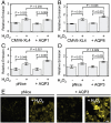
Fig. 1.
Fig. 1.
Application of PY1-ME to show some aquaporins can increase the uptake of H2O2 in mammalian cells. HEK 293 cells transfected with either a control vector or AQP1 (A), AQP8 (B), or AQP3 (C) expression vectors, loaded with PY1-ME, treated with 0 or 50 μM H2O2 for 30 min, and analyzed by flow cytometry (n = 3). (D) HEK 293 cells transfected with either a control vector or AQP3 and mRFP as a transfection marker and measured by confocal microscopy (n = 8). (E) Representative images of experiment represented in (D). Scale bar = 20 μm. For all panels data were normalized to controls and statistical analyses were performed with a one-tailed Student’s t-test. Error bars are ± s.e.m.
Fig. 2.
Fig. 2.
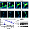
AQP3 mediated exogenous H2O2 uptake can effect intracellular signaling. (A) Live-cell imaging of changes in HyPer fluorescence upon treatment of HEK 293 cells with 10 μM H2O2. “pNice,” HEK 293 cells expressing HyPer and transfected with pNice as a control vector before addition of H2O2. “+AQP3,” HEK 293 cells expressing HyPer and AQP3 before addition of H2O2. “+H2O2,” cells 2 min after addition of 10 μM H2O2. 20 μm scale bar. (B) Live-cell imaging of changes in HyPer fluorescence upon treatment of HeLa cells with 10 μM H2O2. “pNice,” HeLa cells expressing HyPer and transfected with pNice as a control vector before addition of H2O2. “+AQP3,” HeLa cells expressing HyPer and AQP3 before addition of H2O2. “+H2O2,” cells 2 min after addition of 10 μM H2O2. 20 μm scale bar. (C) Time-course and quantification of (B). HeLa cells expressing HyPer and transfected with pNice (black line) or AQP3 (blue line) were stimulated with 10 μM H2O2 at t = 0 and the changes in HyPer fluorescence monitored over time. Error bars are ± s.e.m. (n = 3). (D) Western blot showing changes in pAKT1/2/3 levels in HeLa cells treated with H2O2. HeLa cells expressing AQP3 or control vector were serum-starved and then treated with 200 μM H2O2 for 20 min at 37 °C and then lysed. Phospho-AKT and AQP3 levels were measured by Western blot analysis of whole cell extracts, followed by stripping and reprobing for total AKT or Actin, respectively.
Fig. 3.
Fig. 3.
Natural levels of AQP3 can mediate exogenous H2O2 uptake. (A) HT29s transfected with either HyPer and shRNA targeted against AQP3(241) or AQP4(801) as a control with 100 μM H2O2 at t = 0 and the changes in HyPer fluorescence monitored over time. Images show cells at t = 0 and t = 2 min (“+H2O2”). 10 μm scale bar. (B) Knockdown of endogenous AQP3 in HT29 cells. HT29 cells were transfected with either shRNA targeted against AQP3(241), or AQP4(801) as a control, then lysed and probed for AQP3 levels by Western blot analysis of whole cell extracts, followed by stripping and reprobing for Actin. (C) Endogenous AQP3 can mediate H2O2 uptake. HT29 cells expressing HyPer and transfected with AQP4 shRNA as a control (black line) or AQP3 shRNA (blue line) were stimulated with 100 μM H2O2 at t = 0 and the changes in HyPer fluorescence monitored over time. Error bars are ± s.e.m. (n = 3). (D) Quantification of C at t = 2. (n = 3). Data were normalized to controls and statistical analyses were performed with a one-tailed Student’s t test. Error bars are ± s.e.m.
Fig. 4.
Fig. 4.
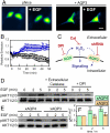
Natural levels of AQP3 can mediate uptake of Nox-generated H2O2 and effect intracellular signaling. (A) Live-cell imaging of changes in HyPer fluorescence upon treatment of HT29 cells with 100 ng/mL EGF. HT29 cells expressing HyPer and transfected with pNice as a control (“pNice” ) or AQP3 (“+AQP3”) before addition of EGF. Cells images at t = 0 and then again at t = 20 min (“+EGF”). 20 μm scale bar. (B) Time-course and quantification of A. HT29 cells expressing HyPer and transfected with pNice (black line) or AQP3 (blue line) stimulated with 100 ng/mL EGF at t = 0 and the changes in HyPer fluorescence monitored over time. Error bars are ± s.e.m. (n = 3). (C) Schematic illustrating where the H2O2 redox signal can by intercepted. EGF stimulation can activate a membrane-bound NADPH oxidase (Nox), which will then produce extracellular H2O2, which can then pass into the cell and modulate intracellular redox signaling. Diphenylene iodonium (DPI) will block the Nox production of H2O2 and extracellular catalase (Cat) will destroy any H2O2 produced outside of the cell. The genetic manipulation of AQP3 using shRNA will block any potential factilitated uptake of extracellular H2O2 by this protein. (D) Extracellular catalase and DPI abrogate EGF signaling in HT29 cells. Serum-starved HT29 cells were either preincubated with 5 μM DPI or DMSO as a carrier control for 30 min. 5 mg/mL catalase or water as a carrier control was then added while the cells were simulataneously stimulated with 100 ng/mL EGF. Phospho-AKT was measured by Western blot analysis of whole cell extracts, followed by stripping and reprobing for total AKT. (E) HT29 cells were transfected with either AQP3 shRNA or AQP4 shRNA as a control, serum starved, stimulated with 100 ng/mL EGF, and lysed at the various time points. Phospho-AKT was measured by Western blot analysis of whole cell extracts, followed by stripping and reprobing for total AKT. (F) Quantification of experiment as represented in (E) by analyzing 4 separate trials. pAKT normalized to total AKT and each experiment normalized to the t = 0 pAKT/AKT ratio. Error bars are ± s.e.m.

Similar articles

Cited by

References

    1. Poole LB, Karplus PA, Claiborne A. Protein sulfenic acids in redox signaling. Annu Rev Pharmacol Toxicol. 2004;44:325–347. - PubMed
    1. Rhee SG. Cell signaling H2O2, a necessary evil for cell signaling. Science. 2006;312:1882–1883. - PubMed
    1. D’Autreaux B, Toledano MB. ROS as signalling molecules: mechanisms that generate specificity in ROS homeostasis. Nat Rev Mol Cell Biol. 2007;8:813–824. - PubMed
    1. Winterbourn CC. Reconciling the chemistry and biology of reactive oxygen species. Nat Chem Biol. 2008;4:278–286. - PubMed
    1. Paulsen CE, Carroll KS. Orchestrating redox signaling networks through regulatory cysteine switches. ACS Chem Biol. 2010;5:47–62. - PMC - PubMed

Publication types